If you or a loved one has been diagnosed with a complex retinal condition, the term vitreoretinal surgery might feel overwhelming. Whether you are dealing with a retinal detachment, macular hole, or complications from diabetic retinopathy, understanding the process is the first step toward a successful recovery.
At Retina and Vitreous Surgeons of Utah (RVSU), we believe that an informed patient is a confident patient. Our goal is to provide world-class surgical care while ensuring you feel supported at every stage of your journey. This guide outlines exactly what you can expect before, during, and after your procedure.
What is Vitreoretinal Surgery?
Vitreoretinal surgery refers to a group of specialized procedures performed in the deep interior of the eye, specifically involving the vitreous gel and the retina. The most common form of this surgery is a vitrectomy, where the surgeon removes the vitreous humor—the clear gel that fills the eye—to gain access to the retina for repairs.
This delicate surgery is used to treat several sight-threatening conditions, including:
- Retinal Detachment: Reattaching the light-sensitive tissue at the back of the eye.
- Macular Pucker or Hole: Repairing the central part of the retina responsible for sharp vision.
- Vitreous Hemorrhage: Removing blood that has leaked into the vitreous gel.
- Diabetic Retinopathy: Addressing scar tissue or leaking vessels caused by diabetes.
Before Vitreoretinal Surgery: Preparing for Your Procedure
The road to a successful outcome begins long before you enter the operating room. Once your RVSU surgeon determines that vitreoretinal surgery is the best course of action, our team will guide you through the preparation phase.
Medical Evaluation
You may need a “clearance” exam from your primary care physician to ensure you are healthy enough for the procedure. Your surgeon will also review your current medications, particularly blood thinners, which may need to be adjusted before surgery.
Pre-Operative Instructions
Typically, you will be asked to:
- Fast (nothing to eat or drink) after midnight the night before surgery.
- Arrange for a family member or friend to drive you home, as you cannot drive yourself after receiving sedation.
- Prepare your home for recovery, ensuring you have easy-to-prepare meals and a comfortable place to rest.
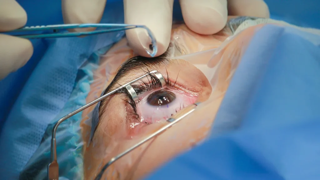
During Vitreoretinal Surgery: What Happens in the Operating Room?
Many patients worry about being awake or feeling pain during eye surgery. At Retina and Vitreous Surgeons of Utah, we prioritize patient comfort using modern anesthesia techniques.
Anesthesia and Comfort
Most vitreoretinal procedures are performed on an outpatient basis. You will likely receive “monitored anesthesia care,” which involves local anesthesia to numb the eye completely and intravenous sedation to keep you relaxed and “twilight” sleepy. You will not feel pain during the procedure.
The Procedure
Using a high-powered operating microscope and micro-incisional tools (often no thicker than a human hair), your surgeon will:
- Make tiny incisions in the white part of the eye (sclera).
- Remove the vitreous gel.
- Perform the necessary repairs (using lasers to seal tears or forceps to remove scar tissue).
- Replace the vitreous with a substitute, such as saline, a gas bubble, or silicone oil, to hold the retina in place.
Because the incisions are so small, they are often “self-healing,” meaning stitches are frequently not required.
After Vitreoretinal Surgery: The Path to Recovery
The postoperative phase is the most critical period for ensuring the retina heals correctly. While every patient’s recovery timeline is unique, there are several universal expectations.
Immediate Aftercare for Vitreoretinal Surgery
You will leave the surgery center with a protective shield over your eye. It is normal to experience some mild “grittiness,” redness, or swelling. Your surgeon will prescribe a regimen of eye drops—usually a combination of antibiotics and anti-inflammatories—to prevent infection and reduce swelling.
The Importance of Positioning
If a gas bubble was used during your surgery, face-down positioning (or “prone positioning”) may be required. This is perhaps the most challenging part of recovery, but it is vital. The gas bubble acts as a temporary bandage, pressing the retina against the back of the eye so it can reattach. Your surgeon will specify how many days you must maintain this position.
What to Avoid During Recovery
To protect your eye while it heals, you must follow these restrictions:
- No Heavy Lifting: Avoid straining or lifting objects over 10–15 pounds.
- No Air Travel: If a gas bubble was used, you cannot fly or travel to high altitudes (like Utah’s mountain passes) until the bubble has dissipated. Changes in air pressure can cause the bubble to expand, leading to dangerous increases in eye pressure.
- No Eye Rubbing: Protect the eye from any direct pressure.
When to Call Your Surgeon
While complications are rare, you should contact Retina and Vitreous Surgeons of Utah immediately if you experience:
- A sudden decrease in vision.
- Severe pain that is not relieved by over-the-counter medication.
- An increase in new “floaters” or flashes of light.
Why Choose Retina and Vitreous Surgeons of Utah (RVSU)?
When it comes to your vision, experience matters. Our surgeons are board-certified and fellowship-trained in the most advanced vitreoretinal techniques. We combine cutting-edge technology with a compassionate, patient-centered approach to ensure you receive the highest standard of care in the Intermountain West.
Vitreoretinal surgery is a journey, and we are with you every step of the way—from the initial diagnosis to the final follow-up.