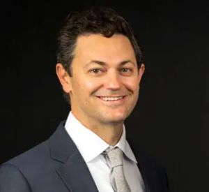
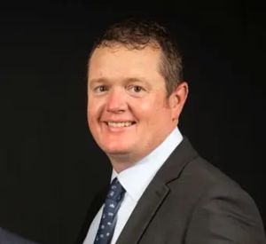
A retinal occlusion happens when a blood vessel in the back of your eye becomes blocked. Our board-certified retina specialists at RVSU diagnose and treat all types of vascular blockages, from early swelling to complex vitreoretinal surgery. All four of our physicians trained at the number one eye hospital in the country.
vascular cause of vision loss after diabetic retinopathy in the United States.
are linked to high blood pressure, making regular health screenings a vital part of care.
worldwide are affected by retinal vein blockages, requiring specialized medical attention.
There are two main types of clogs, depending on which blood vessel is affected:
Fluid collects in the center of the retina. This causes blurring in the vision you use for reading and recognizing faces. Frequent follow-up exams are usually needed.
This occurs when the retina is starved of oxygen because of the clog. Without fresh blood flow, the delicate nerve cells in the eye can begin to stop working.
The eye tries to grow new, weak blood vessels to replace the blocked ones. These vessels bleed easily and can cause high eye pressure or a “curtain” in your vision.
Long-term blockages can lead to scarring or a detached retina. This stage requires urgent surgical treatment to prevent total blindness and stabilize the eye.
An eye stroke usually happens suddenly and almost always in only one eye. Contact our office right away if you notice any of the following:
Notice box: If you lose your vision suddenly, do not wait. This is a medical emergency. A retina specialist can check your eye and start treatment to try and save your vision.
At every visit, our specialists perform a thorough dilated eye exam. We use the most advanced imaging available to identify the exact location of the blockage and create a treatment plan that fits your needs.
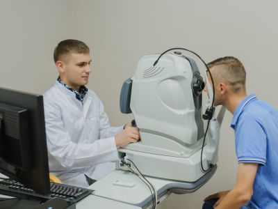
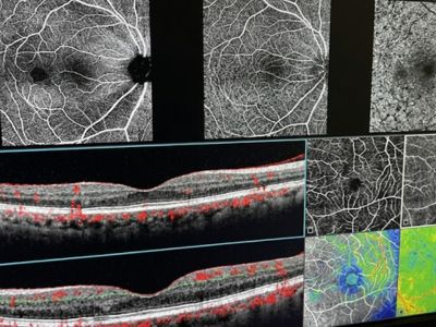
This noninvasive imaging test uses light waves to create detailed retinal maps, helping doctors detect fluid, swelling, and subtle changes.
During this test, dye injected into your arm travels to eye vessels, allowing imaging to identify leaks, blockages, or growth.
OCTA is a dye free imaging method maps retinal blood flow quickly, providing insights into circulation and detecting early changes.
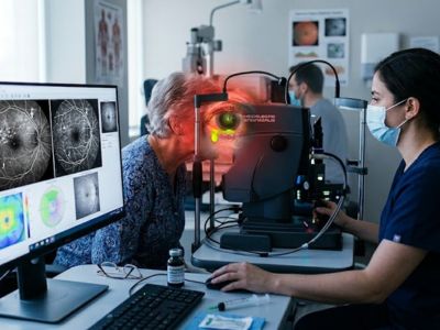
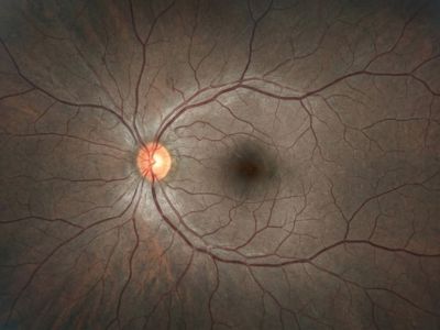
We use specialized cameras to capture high resolution images of your retina, creating baseline records and tracking even small changes.
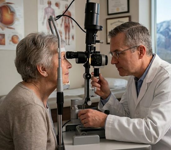
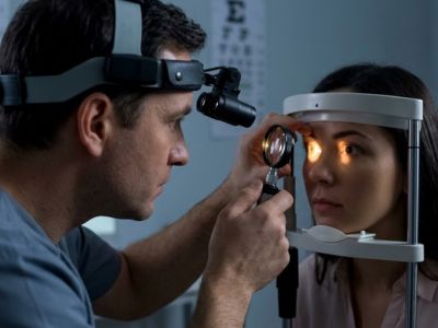
During this exam, doctors dilate your pupils and examine retina, vitreous, and optic nerve to ensure no detail vision missed.
Anti-VEGF medications are injected into the eye to stop abnormal blood vessel growth and reduce leakage. This is the first-line treatment for swelling caused by a vein occlusion. We use Eylea, Lucentis and Avastin. Injections typically start monthly and are spaced out as your condition improves.
Laser treatment seals leaking blood vessels and reduces the signal that drives new vessel growth. This prevents dangerous bleeding inside the eye and helps lower eye pressure. Treatment is done in our office over one to three sessions and is often combined with anti-VEGF injections.
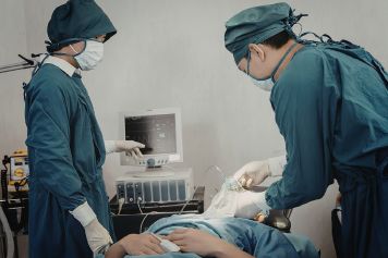
For advanced cases where bleeding has entered the vitreous or the retina has detached, surgery is needed. Our surgeons perform vitrectomy to clear blood from the eye, release traction on the retina, and repair detachments. RVSU performs over 100 retina surgeries per month.

All four of our physicians are board-certified ophthalmologists with fellowship training in vitreoretinal surgery. Retinal vascular disease is a clinical focus for each of them.
Sudden vision changes are a sign that your eye needs expert care. We are accepting new patients at four locations across Utah. Referrals from primary care physicians and cardiologists are welcome.
See all of our convenient locations across Utah.