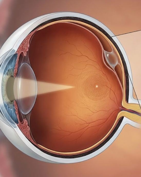
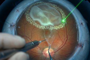
Retinoschisis is a condition where the layers of your retina begin to split apart. This creates a small fluid-filled area that can look very similar to a retinal detachment. Our board-certified retina specialists at RVSU use high-tech imaging to tell the difference and monitor your eye health.
Most cases of retinoschisis are found during a routine eye exam and do not cause any symptoms. However, if the splitting becomes severe, it can lead to vision loss or a full detachment. Because a professional diagnosis is so important, we use the latest tools to provide a clear answer for your retinal splitting symptoms.
over the age of 40 are estimated to have some form of degenerative retinoschisis.
of retinoschisis may lead to a progressive retinal detachment that requires surgery.
with this condition have it in both eyes, requiring a complete exam of the entire retina.
Retinoschisis happens when the delicate nerve tissue of the retina splits into two separate layers. This often occurs because of aging or a genetic trait. While the split layers usually stay in place, they can sometimes cause a “blister” to form in your side vision.
Degenerative Retinoschisis
This is the most common type and is usually linked to aging. It typically affects the far edges of the eye and does not impact your central vision. Most people never notice a change in their sight and only find out about it during a dilated exam.
Juvenile Retinoschisis
This is a rare, genetic form that affects young boys. It often impacts the center of the retina (the macula). This can cause blurring or a loss of detailed vision early in life, making early eye exams vital for children with a family history.
Know Your Risk
You are at a higher risk if you are extremely nearsighted or have a family history of retinal issues. Regular checkups are the only way to ensure the split does not grow larger or allow fluid to leak into dangerous areas.

The split starts in the far corners of the eye. There is no risk to your central vision, and most patients have no symptoms. Annual monitoring is the standard of care.
The area of the split grows larger and becomes more elevated. You may notice a slight decrease in your side vision. More frequent exams are needed to track these changes.
Holes begin to form in the inner layer of the split retina. This increases the risk of fluid moving through the tissue. Our specialists watch these holes closely for any signs of instability.
Holes form in both the inner and outer layers. This allows fluid to get behind the retina, which can cause a full detachment and requires urgent medical or surgical care.
Because retinoschisis usually happens in the side vision, most people have no symptoms. However, contact our office right away if you notice:
Notice box: Retinoschisis can look exactly like a retinal detachment to an untrained eye. A fellowship-trained retina specialist is needed to provide the correct diagnosis and prevent unnecessary surgery.
At every visit, our specialists perform a thorough dilated eye exam. We use the most advanced imaging available to identify the exact layers of the split.
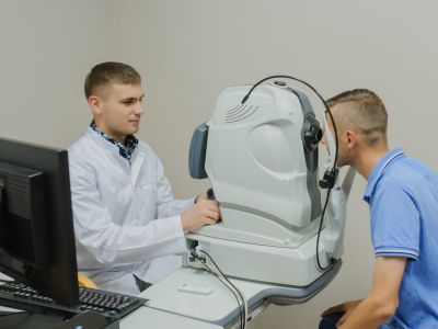
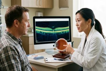
This non-invasive test is the most important tool for retinoschisis. It produces a detailed map that shows the doctor exactly where the retina is splitting and how thick each layer is.
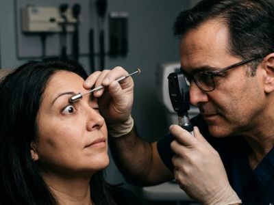
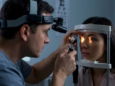
The doctor uses a small tool to gently move the eye during the exam. This allows them to see the far peripheral retina more clearly and check for any tiny holes that could lead to a detachment.
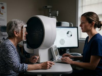
This test measures your side vision to see if the retinoschisis is causing any “blind spots.” This helps us understand how much the condition is affecting your everyday sight.
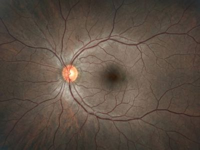
We use specialized cameras to capture the far edges of the retina. These photos allow us to track the size of the “blister” over time and ensure it is not moving toward the center of your eye.
Every appointment includes a hands-on clinical exam. Our fellowship-trained physicians personally examine your retina to ensure the tissue is healthy and the condition is stable.
Most patients with retinoschisis do not need active treatment. Our specialists will review your imaging and create a plan based on the stability of your retina.
If the split is stable and far from the center of the eye, we monitor it once a year. We will provide you with a list of symptoms to watch for so you know when to call for an urgent checkup.
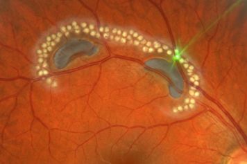
If holes form in the retina or fluid starts to move, we may use a laser to “barrier” the area. This creates a seal that prevents the split from turning into a full retinal detachment.
In rare cases where a detachment occurs, surgery is required. Our surgeons perform a vitrectomy or scleral buckle to repair the retina and protect your sight. RVSU performs over 100 retina surgeries per month.
All four of our physicians are board-certified ophthalmologists with fellowship training in vitreoretinal surgery. Managing complex retinal splitting is a clinical focus for each of them.
Proper diagnosis is the most important part of managing retinoschisis. We are accepting new patients at four locations across Utah. Referrals from primary care physicians and optometrists are welcome.
See all of our convenient locations across Utah.