
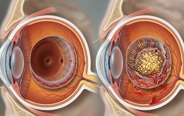
Many adults notice changes in their central vision as they age, and macular degeneration is one of the most common reasons for those changes. Age-related macular degeneration (AMD) is the leading cause of permanent vision loss in adults over 50 in the United States. Yet millions of people do not catch it until real damage has already been done.
Understanding AMD begins with knowing how it affects the retina, the factors that increase risk, and the treatment options available today.
What Is Macular Degeneration?
Your eye has a small, highly sensitive area at the centre of the retina called the macula. It is responsible for sharp, detailed, straight-ahead vision, the kind you need to read, drive, recognise faces, and see fine details.
Macular degeneration is the gradual breakdown of this tissue. As the macula deteriorates, central vision becomes blurry, distorted, or entirely lost. Peripheral (side) vision usually stays intact, but the loss of central sight profoundly affects daily quality of life.
There are two main forms of AMD:
- Dry AMD (Non-Neovascular): This is the more common form of the condition. It develops when tiny yellow deposits called drusen build up beneath the retina, slowly damaging macula cells over time. Dry AMD progresses gradually and may not cause noticeable symptoms in early stages, but in advanced cases, it can develop into geographic atrophy, a serious loss of retinal tissue.
- Wet AMD (Neovascular): Though less common, wet AMD is responsible for approximately 90% of severe vision loss from AMD. It occurs when abnormal blood vessels grow beneath the retina and leak fluid or blood, causing rapid and significant damage to central vision. Wet AMD can progress within days or weeks, making early detection critical.
What Causes Macular Degeneration?
AMD does not have a single cause. It develops from a complex combination of age, genetics, and everyday lifestyle choices.
| Risk Factor | Details |
|---|---|
| Age | Risk increases significantly after age 50; most cases occur in older adults. |
| Genetics & Family History | Having a first-degree relative with AMD raises your risk considerably. |
| Smoking | Recognized as a primary modifiable risk factor that increases development rates. |
| Cardiovascular Disease | High blood pressure and high cholesterol are linked to AMD progression. |
| UV Exposure | Long-term unprotected sun exposure may contribute to retinal damage. |
| Race | Caucasians are at higher risk than other racial groups. |
| Obesity | A high BMI, particularly with a sedentary lifestyle, increases AMD risk. |
| Diet | Low intake of antioxidant-rich foods is associated with faster progression. |
Knowing which risk factors apply to you can help you make informed decisions about your eye health and regular screenings.
Symptoms of Macular Degeneration
In many cases, early dry AMD develops gradually and may not cause obvious vision changes at first. Some patients do not realise they have AMD until routine testing reveals retinal changes. Watch for these warning signs:
- Blurry or fuzzy central vision that does not improve with glasses. This is often one of the first things people notice: text on a page looks smudged or faces seem unclear even at normal distances.
- Straight lines appear wavy or bent. This is called metamorphopsia and is a hallmark symptom of wet AMD. If the lines on a printed page or window blinds suddenly look curved, seek care immediately.
- A dark, blurry, or empty spot in the centre of your visual field. This scotoma (blind spot) can grow over time with untreated AMD.
- Colours appear washed out or less vivid. The macula processes colour detail, and as it degenerates, colour perception dulls.
- Difficulty adapting from bright to dim light, making activities like driving at dusk or night increasingly difficult.
One simple at-home monitoring tool is the Amsler Grid, a grid of straight lines with a dot in the centre. Covering one eye at a time, look at the dot. If the surrounding lines appear wavy, missing, or distorted, you should contact a retina specialist right away.
How Is Macular Degeneration Diagnosed?
A comprehensive dilated eye exam is the foundation of AMD diagnosis. At a specialised retina practice like Retina and Vitreous Surgeons of Utah (RVSU), the diagnostic workup typically includes the following:
- Fluorescein Angiography: A specialised dye is injected into the bloodstream, and rapid photographs track its flow through the retinal blood vessels, revealing leaking or abnormal vessel weaknesses associated with wet AMD.
- Optical Coherence Tomography (OCT): This non-invasive imaging scan produces cross-sectional images of the retina, allowing doctors to measure retinal thickness, detect fluid beneath the macula, and monitor changes over time with exceptional detail.
- Colour Fundus Photography: High-resolution photos of the retina document the presence and severity of drusen and other AMD-related changes, creating a visual baseline for tracking progression.
The location of any neovascular (abnormal vessel) lesions, whether subfoveal (directly under the fovea) or non-subfoveal, plays a major role in determining your specific treatment approach and overall prognosis.
Macular Degeneration Treatment Options
Treatment Approaches for Dry AMD
Currently, no medical treatment reverses dry AMD. However, the landmark AREDS and AREDS2 clinical trials found that high-dose antioxidant supplements, specifically vitamin C, vitamin E, lutein, zeaxanthin, zinc, and copper, can reduce the risk of progression to advanced stages in people with intermediate to advanced dry AMD.
Lifestyle habits also play an important role in supporting long-term retinal health: quitting smoking, maintaining healthy blood pressure and cholesterol, eating a diet rich in leafy greens and omega-3 fatty acids, and wearing UV-protective eyewear are all evidence-based strategies to slow progression.
Medical Management of Wet AMD
Wet AMD requires prompt, active treatment. Advances in retinal medicine have significantly improved treatment outcomes for many patients with wet AMD.
- Anti-VEGF Injections (Intravitreal Injections): This is the gold-standard treatment for wet AMD. Medications such as ranibizumab (Lucentis), bevacizumab (Avastin), and aflibercept (Eylea) are injected directly into the eye to block vascular endothelial growth factor (VEGF), the protein that drives abnormal blood vessel growth. Most patients require monthly injections initially, with treatment frequency adjusted based on response. Studies show that anti-VEGF therapy not only stops vision loss in most patients but actually improves vision in a significant portion of cases.
- Photodynamic Therapy (PDT): This approach involves intravenous administration of verteporfin (Visudyne), a light-activated drug that concentrates in abnormal retinal blood vessels. A non-thermal laser is then applied to activate the drug, selectively destroying the abnormal vessels without damaging surrounding tissue. PDT is typically repeated every three months as needed. Patients must avoid direct sunlight and bright indoor light for five days following treatment.
- Thermal Laser Photocoagulation: Used selectively for specific AMD lesion types located away from the centre of the macula, thermal laser destroys abnormal blood vessels directly. Because it can cause scarring, its use is more limited in modern AMD management.
Comparing Dry and Wet AMD
| Feature | Dry AMD | Wet AMD |
|---|---|---|
| Prevalence | Most common form of AMD | Less common form of AMD |
| Cause | Drusen deposits, slow cell death | Abnormal blood vessel growth under the retina |
| Progression | Slow, over years | Rapid, within weeks or days |
| Vision Loss | Gradual central vision blurring | Severe central vision loss, can be sudden |
| Primary Treatment | AREDS study supplements, lifestyle | Anti-VEGF injections, PDT options |
| Reversible? | Not currently | Partially, with early treatment intervention |
Can You Prevent Macular Degeneration?
You cannot change your age or genetics, but you can meaningfully reduce your risk and slow progression:
- Eat a Mediterranean-style diet loaded with dark leafy greens, colourful vegetables, fish, and nuts. These foods provide the lutein, zeaxanthin, and omega-3 fatty acids that support retinal health.
- Stop smoking — it is the single most modifiable risk factor for AMD.
- Protect your eyes with UV-blocking sunglasses outdoors.
- Keep blood pressure and cholesterol in a healthy range.
- Get regular dilated eye exams after age 50, or sooner if you have a family history.
Conclusion
Facing a diagnosis or dealing with the warning signs of macular degeneration can feel overwhelming, but it is important to remember that you have actionable choices. While dry AMD requires diligent daily tracking and lifestyle adjustments, modern medical treatments for wet AMD are incredibly effective at protecting and sometimes even improving your sight. The difference between catching AMD early and catching it late can be the difference between preserving functional vision and losing the ability to read, drive, or recognise the faces of people you love.
At Retina and Vitreous Surgeons of Utah (RVSU), board-certified retinal ophthalmologists fellowship-trained at the nation’s top eye institutions offer expert diagnosis and the full spectrum of AMD treatments across locations in Murray, Draper, Provo, and Payson. By monitoring your eyes at home with the Amsler Grid, staying consistent with eye exams, and acting quickly if your vision shifts, you can preserve your independence and continue seeing the world clearly. If you or someone you care about is experiencing changes in central vision or is simply overdue for a retina evaluation, please contact us today to schedule a comprehensive evaluation and protect your sight.