
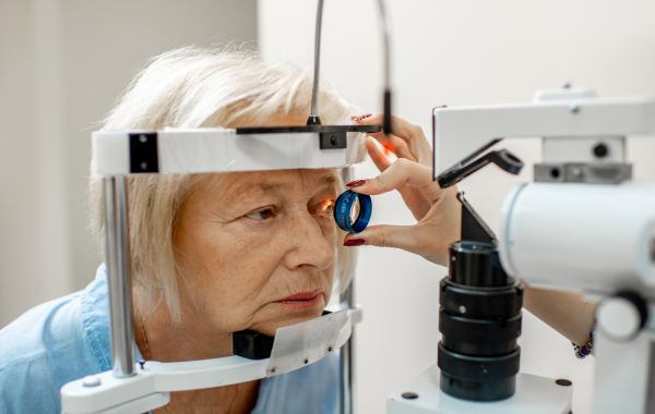
Many people think glaucoma and retinal disease are two different eye problems. But they can happen together and affect your vision at the same time. Understanding how glaucoma and retinal conditions intersect can help you protect your eyesight before small problems turn into big ones.
We see patients every week who come in for one eye problem and later learn a second one is quietly developing. This blog breaks down the connection in plain, simple language, so you know what to watch for and when to see a specialist.
What Is Glaucoma?
Glaucoma is a group of eye diseases that damage the optic nerve. The optic nerve sends pictures from your eye to your brain. Glaucoma often happens when fluid inside the eye cannot drain properly. This raises the pressure inside the eye, called eye pressure or IOP.
Over time, this pressure can slowly destroy the tiny nerve fibers called retinal ganglion cells. These cells do not grow back once they’re gone. That’s why glaucoma is often called the silent thief of sight. Most people don’t notice any symptoms until they’ve already lost a good amount of side vision.
What Are Retinal Conditions?
The retina is the thin layer of tissue lining the back of your eye. You can think of the retina as the part of the eye that captures light and helps your brain create images. When the retina gets damaged, your vision suffers directly.
Common retinal conditions include:
- Diabetic retinopathy damage to blood vessels in the retina caused by high blood sugar
- Age-related macular degeneration (AMD) breakdown of the macula, the part of the retina responsible for sharp, central vision
- Retinal detachment when the retina pulls away from the back wall of the eye
- Uveitis inflammation inside the eye that can affect the retina and other structures
- Retinal vein occlusion a blockage in the veins that carry blood away from the retina
Where Glaucoma and Retinal Disease Overlap
Here’s the part most people don’t know: these two conditions can cause each other, hide each other, and make each other worse. Let’s break down exactly how.
1. Retinal Disease Can Cause Glaucoma
- Abnormal Blood Vessels: Conditions like diabetic retinopathy can trigger fragile, abnormal blood vessels to grow inside the eye.
- Drainage Blockage: These blood vessels can block the eye’s drainage system and quickly raise eye pressure.
- Neovascular Glaucoma: This can lead to a serious type of glaucoma that needs treatment right away.
2. Glaucoma Directly Affects the Retina
- Ganglion Cell Damage: Glaucoma thins the specific layers of the retina where crucial ganglion cell axons reside.
- OCT Tracking: Doctors rely on retinal OCT (optical coherence tomography) scans to measure this thinning layer by layer.
- Disease Progression: Tracking retinal thinning gives specialists a clear picture of how far glaucoma has advanced.
3. Shared Risk Factors
- Cardiovascular Issues: High blood pressure, poor blood flow, and other health problems can increase your risk of both conditions.
- Metabolic & Vascular Conditions: Living with diabetes, chronic migraines, or sleep apnea elevates risk across the board.
- Dual Screening: If you have any of these conditions, getting both your retina and optic nerve checked regularly is essential.
4. One Condition Can Mask the Other
- Diagnostic Confusion: When both conditions occur together, pinpointing which one is causing vision loss becomes difficult.
- Skewed Test Results: Retinal swelling or macular damage can alter the results of standard visual field tests used to track glaucoma.
- Comprehensive Care: An experienced team must evaluate the full clinical picture rather than relying on a single test.
5. Having Both Amplifies Vision Loss Risk
- Compounded Effects: Overlapping conditions like glaucoma alongside macular degeneration or diabetic retinopathy accelerate vision degradation.
- Higher Blindness Risk: People with both conditions have a higher risk of serious vision loss.
- Early Intervention: Finding and treating both conditions early can help protect your vision
Quick Comparison: Glaucoma vs. Retinal Disease
| Feature | Glaucoma | Retinal Disease |
|---|---|---|
| Main area affected | Optic nerve | Retina (back layer of the eye) |
| Common cause | High eye pressure | Blood vessel damage, aging, inflammation, injury |
| Vision loss pattern | Side (peripheral) vision first | Central vision first (varies by type) |
| Early symptoms | Usually none | Blurry spots, wavy lines, flashes of light |
| Main test used | Eye pressure check, visual field test | OCT scan, dilated eye exam, retinal imaging |
| Reversible? | No, damage is permanent | Sometimes, if caught early |
| Can they occur together? | Yes | Yes |
Symptoms You Shouldn't Ignore
Both conditions may not cause symptoms at first. Watch for these warning signs and see an eye doctor if you notice them:
- Blurry or wavy central vision
- Trouble seeing in low light or at night
- Dark spots or gaps in your side vision
- Sudden flashes of light or new floaters
- A curtain-like shadow moving across your vision
- Colors looking faded or washed out
- Needing brighter light to read
If you notice any of these, don’t wait. Sudden changes, especially flashes, floaters, or a shadow in your vision, should be treated as an emergency.
Who Is Most at Risk?
Some people have a higher chance of developing both conditions. You may be at greater risk if you:
- Are over the age of 40
- Have diabetes
- Have high blood pressure or poor circulation
- Have a family history of glaucoma or retinal disease
- Are highly nearsighted (high myopia)
- Have had an eye injury or previous eye surgery
- Smoke
If your parents or grandparents had retinal disease or glaucoma, your own risk for these conditions goes up. Knowing your family history is one of the simplest ways to stay ahead of eye disease.
How Eye Doctors Diagnose Both Conditions
A thorough eye exam usually checks for both glaucoma and retinal problems at the same time, since they overlap so often. Here’s what that typically includes:
- Dilated eye exam drops widen your pupil so the doctor can see the retina and optic nerve clearly
- Tonometry measures the pressure inside your eye
- OCT imaging creates a detailed cross-section picture of the retina and optic nerve
- Visual field test checks for blind spots or missing areas in your side vision
- Fundus photography takes a picture of the back of the eye to track changes over time
Sometimes retinal disease can affect glaucoma test results. Your eye doctor and retina specialist may work together to plan your care. This teamwork is often the key to getting an accurate diagnosis.
How These Conditions Are Treated Together
Treatment depends on which condition is more advanced and how much they’re affecting each other. Some common approaches include:
- Eye pressure-lowering drops or laser treatment for glaucoma
- Injections into the eye to treat swelling or abnormal blood vessels from retinal disease
- Laser therapy to seal leaking blood vessels or repair retinal tears
- Surgery in more advanced cases of either condition
- Managing underlying health issues, like blood sugar or blood pressure, to slow down both diseases
The goal is always the same protect as much vision as possible and stop further damage before it happens.
Protecting Your Vision Going Forward
You can’t always prevent glaucoma or retinal disease, but you can lower your risk and catch problems early. Here are simple habits that make a real difference:
- Get a full dilated eye exam every one to two years, more often if you’re over 40 or have risk factors
- Keep blood sugar and blood pressure under control
- Don’t skip follow-up appointments, even if your vision feels fine
- Wear protective eyewear during sports or risky activities
- Tell your eye doctor about your family history
- Report any sudden vision changes right away
When to See a Retina Specialist
If you already have glaucoma, or a family history of it, and you notice any changes in your central vision, it’s time to see a retina specialist along with your regular eye doctor. Catching a retinal problem early, before it complicates your glaucoma treatment, gives you the best chance of keeping your sight for years to come.
Final Thoughts
Glaucoma and retinal conditions don’t just happen to exist in the same eye by chance. They share risk factors, can directly trigger one another, and often complicate diagnosis when present at the same time. Because symptoms can develop quietly, staying proactive with regular dilated eye exams and paying close attention to any sudden vision changes is your best line of defense.
If you have concerns about your eye pressure, retinal health, or are experiencing changes in your sight, early evaluation is essential. Please contact us today to schedule an appointment with our specialist team and take the first step toward protecting your vision long-term.