
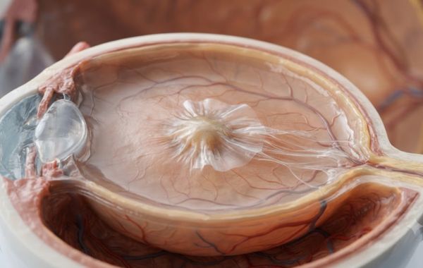
If your central vision has started to look a bit wavy or blurry lately, you might be dealing with something called a macular pucker. It’s a fairly common eye condition, and most people have never heard the term until an eye doctor brings it up during a routine exam. The condition sounds unusual, but it’s actually pretty easy to understand once you know what’s happening inside the eye.
A macular pucker develops slowly, often without any warning signs at first, and it shares some traits with other retinal conditions like macular degeneration. Below, we’ll go over what causes it, what it does to your eyesight, and what an eye doctor can do about it.
What Is a Macular Pucker?
A macular pucker is a thin layer of scar tissue that forms on your macula. The macula is the small central part of your retina, and it’s the part responsible for your sharp, detailed vision. You use it every time you read fine print or recognize a face from across the room.
Doctors also call this condition an epiretinal membrane, or sometimes cellophane maculopathy, because the tissue looks a bit like a thin sheet of cellophane wrap sitting on the retina. When this scar tissue tightens or shrinks, it pulls on the macula underneath it. That pulling causes the smooth surface of the macula to wrinkle, or “pucker.” It’s this wrinkling that leads to the vision changes people notice.
Why Does a Macular Pucker Happen?
Most macular puckers happen simply because of age. Inside your eye, there’s a gel-like substance called the vitreous that fills most of the space between your lens and your retina. As you get older, this gel slowly shrinks and starts to pull away from the retina. This is a normal process called posterior vitreous detachment, and most people go through it without any problems at all.
But sometimes, as the vitreous pulls away, tiny bits of it stay stuck to the retina’s surface. This leaves behind microscopic damage. Your retina then tries to heal itself, and in doing so, it forms a thin layer of scar tissue over the area. That scar tissue is the macular pucker. It’s a bit like how a scab forms on skin after a scrape, except this scab forms on the delicate surface of your retina instead.
Common Causes and Risk Factors
A macular pucker can form on its own with no clear reason, and this is actually the most common situation. But there are also some conditions and events that raise your risk.
- Age: Most cases happen in people over 50, and the risk keeps climbing after that.
- Posterior vitreous detachment: This natural aging process is the leading trigger behind most macular puckers.
- Retinal tears or detachment: Damage to the retina can prompt scar tissue to form as part of the healing process, similar to what happens with retinal detachment.
- Eye surgery: Procedures like cataract surgery can sometimes lead to inflammation that triggers a pucker afterward.
- Diabetes: People with diabetic eye disease have a higher chance of developing a macular pucker.
- Eye trauma or injury: A blow to the eye can damage the retinal surface and set off the same healing response.
- Eye inflammation or infection: Conditions that inflame the inside of the eye can also be a factor.
How Does a Macular Pucker Affect Vision?
The macula controls your central, detailed vision, so that’s exactly where the changes show up. Your side, or peripheral, vision stays completely normal, which separates macular pucker from several other eye diseases that affect the whole visual field.
Typical vision changes include:
- Straight lines, like door frames or lines on a page, start to look wavy or bent
- Blurry or fuzzy central vision
- Trouble reading small print or fine details
- A gray, cloudy, or blank spot in the middle of your vision
- Objects appearing slightly smaller or larger than they should (this is called micropsia or macropsia)
- Difficulty recognizing faces from a distance
These symptoms often stay mild for years and barely get in the way of daily life, though some people do notice a more significant drop in vision. Macular pucker usually affects only one eye at a time. It can show up in both eyes, but that’s not common.
How Is a Macular Pucker Diagnosed?
Diagnosing a macular pucker usually starts with a simple dilated eye exam. Your doctor puts drops in your eyes to widen your pupils, which lets them get a clear look at the back of your eye, including the macula, using a special lens.
From there, most retina specialists will order a scan called optical coherence tomography, or OCT for short. This is a painless, quick imaging test that takes cross-section pictures of your retina, almost like a slice-by-slice map. OCT can show the exact thickness of the scar tissue and how much it’s pulling on the macula underneath. Your doctor might also use an Amsler grid, which is a simple checkerboard pattern that helps track whether straight lines look distorted to you. This grid is an easy tool you can even use at home to keep an eye on changes between visits.
Treatment Options for Macular Pucker
Not every macular pucker needs treatment. In fact, many cases are mild enough that doctors simply recommend keeping an eye on things with regular checkups.
| Situation | Typical Approach |
|---|---|
| Mild symptoms, minimal vision changes | Observation with regular eye exams, sometimes a new glasses prescription |
| Symptoms are stable but bothersome | Continued monitoring with OCT scans to watch for progression |
| Vision loss interferes with daily tasks like reading or driving | Surgery called a vitrectomy, with membrane peeling |
| Macular pucker linked to another condition, like swelling | Additional treatment, which may include intravitreal injections for the underlying issue |
When surgery is needed, the procedure is called a vitrectomy. During this surgery, a retina specialist removes the vitreous gel and gently peels away the scar tissue from the surface of the macula. This lets the macula relax back into a smoother, flatter shape over time. Most people notice their vision gradually improves over the weeks and months following surgery, though it can take up to three months or longer to see the full results. Vision doesn’t always return to 100 percent, but many patients see meaningful improvement in clarity and less waviness in their central vision.
What Happens If a Macular Pucker Is Left Untreated?
This is a common worry, and macular pucker usually isn’t an emergency. For most people, vision stays fairly stable over the years, and severe vision loss is uncommon.
Still, it’s worth having your eyes checked regularly if you’ve been diagnosed with a macular pucker. In some cases, the scar tissue slowly tightens further, leading to gradually worsening distortion or blurriness. Waiting too long to treat a pucker that’s already causing real vision loss can make it harder for vision to bounce back fully after surgery, since the macula may develop more permanent changes the longer it stays wrinkled. Regular monitoring catches these changes early, while symptoms are still manageable.
Living With a Macular Pucker
Many people live full, normal lives with a macular pucker and never need surgery at all. Since peripheral vision isn’t affected, everyday activities like walking around your house, recognizing people nearby, and general mobility usually aren’t a problem.
Where people notice the biggest impact is with detailed, close-up tasks. Reading, sewing, working on a computer, or driving at night can all feel a bit harder when central vision is distorted. Simple adjustments, like brighter lighting or a magnifier for small print, can make a real difference for day-to-day comfort. If symptoms do start getting in the way of things you need to do, that’s usually the signal it’s time to talk with your retina specialist about next steps.
A Clear Path Forward for Your Vision
A macular pucker can feel unsettling once you notice your vision changing, especially when straight lines start to look wavy or reading gets harder than it used to be. What’s actually happening is scar tissue gently pulling on your macula, and in most cases, that’s a manageable, well-understood problem. Even when treatment is needed, surgery has a solid track record of improving vision.
If you’ve noticed distortion in your central vision, or it’s simply been a while since your last dilated eye exam, the team at Retina and Vitreous Surgeons of Utah can take a closer look at what’s going on. Reach out to schedule an appointment with one of our retina specialists.
Ready to Talk to a Retina Specialist in Utah
If you have been told you need eye injections and still have questions, talk to your retina doctor before the appointment. Knowing what to expect and having a clear aftercare plan takes a lot of the stress out of the whole process.
The team at Retina and Vitreous Surgeons of Utah serves patients across multiple locations in Utah. If you have concerns about your upcoming procedure or want to understand your treatment options, schedule a consultation with one of our retina specialists.