Diabetic Retinopathy Treatment In Utah

Diabetic retinopathy is the leading cause of vision loss in working-age adults. Our board-certified retina specialists at RVSU diagnose and treat all stages of the disease, from early monitoring to complex vitreoretinal surgery. All four of our physicians trained at the number one eye hospital in the country.

No. 1 cause of

blindness in working-age adults in the US

1 in 3 people

with diabetes has some degree of retinopathy

95% of severe

vision loss is preventable with timely treatment

Diabetic Retinopathy

What is Diabetic Retinopathy

High blood sugar damages tiny blood vessels in your retina. They leak, swell, or grow abnormally. No early symptoms, so yearly dilated eye exams are a must for all diabetics.

Non-Proliferative Diabetic Retinopathy (NPDR)

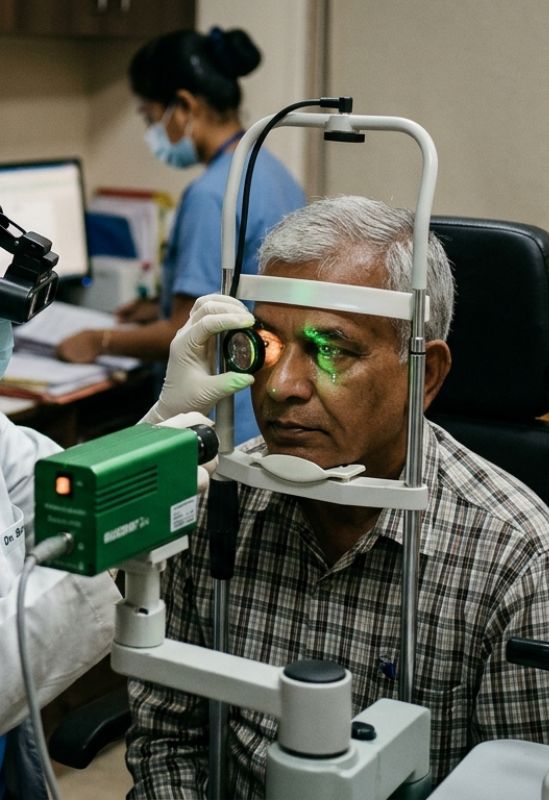
Early stage. Blood vessels weaken and leak. No symptoms at first. Yearly eye exams catch this before it gets worse.

Proliferative diabetic retinopathy (PDR)

Advanced stage. New fragile vessels grow, bleed easily, and can pull the retina loose. See a retina specialist right away.

Diabetic macular edema (DME)

Fluid builds in the macula, blurring the vision you use for reading, driving, and recognizing faces. Most common cause of vision loss.

Know your risk

Long-term diabetes, poor blood sugar, high blood pressure, high cholesterol, or kidney disease all speed up eye damage.

The Four Stages

01

Mild NPDR

Tiny swellings called microaneurysms appear in retinal blood vessels. There are no symptoms. Annual monitoring is the standard of care.

02

Moderate NPDR

More blood vessels become blocked and fluid may begin to collect. Mild blurring can occur. More frequent follow-up exams are needed.

03

Severe NPDR

A large number of blood vessels are blocked. The retina signals the body to grow new vessels. Most people progress to PDR within a year without treatment.

04

Proliferative DR

Abnormal vessels grow on the retina and into the vitreous gel. They bleed easily and can cause retinal detachment. This stage requires urgent treatment.

Symptoms of Diabetic Retinopathy

Diabetic retinopathy often has no symptoms until it reaches an advanced stage. Contact our office right away if you notice any of the following.

Advanced Diagnostics & Imaging

At every visit, our specialists perform a thorough dilated eye exam. We use the most advanced imaging available to identify the exact stage of your condition and create a treatment plan that fits your needs.

Optical Coherence Tomography
A non-invasive imaging test using light waves to create detailed cross-sectional retina images, detecting fluid, swelling, and subtle structural abnormalities early.
Fluorescein Angiography
A diagnostic test where dye is injected into a vein, capturing detailed retinal images to identify leaking, blocked, or abnormal blood vessels accurately.
OCTA (OCT Angiography)
A modern, non-invasive imaging technique that maps retinal blood flow without dye, quickly detecting early vascular changes in a comfortable way.
Wide-Field Fundus Photography
High-resolution imaging captures wide views of the retina, helping establish baseline records and monitor subtle changes in retinal health over time.
Dilated Fundus Exam
A comprehensive eye examination where pupils are dilated, allowing doctors to carefully inspect retina, vitreous, and optic nerve for abnormalities.

Treatments

The right treatment depends on how far the disease has progressed. Our specialists will review your imaging with you and explain all appropriate options before any decisions are made.
Anti-VEGF injections
Anti-VEGF Injections

Anti-VEGF medications are injected into the eye to stop abnormal blood vessel growth and reduce leakage. This is the first-line treatment for diabetic macular edema and proliferative diabetic retinopathy. We use Eylea, Lucentis and Avastin. Injections typically start monthly and are spaced out as your condition improves.

Laser therapy
Laser Therapy
Laser treatment seals leaking blood vessels and reduces the signal that drives new vessel growth. Focal and grid laser is used for macular edema. Panretinal photocoagulation (PRP) is used for proliferative DR. Treatment is done in our office over one to three sessions and is often combined with anti-VEGF injections.
Vitreoretinal surgery
Vitreoretinal Surgery

For advanced cases where bleeding has entered the vitreous or the retina has detached, surgery is needed. Our surgeons perform vitrectomy to clear blood from the eye, release traction on the retina, and repair detachments. RVSU performs over 100 retina surgeries per month.

Our Specialists

All four of our physicians are board-certified ophthalmologists with fellowship training in vitreoretinal surgery.

Schedule Your Appointment Today

Don’t compromise your vision. Schedule an appointment at our Provo office and experience the RVSU difference.

Find an RVSU Location Near You

See all of our convenient locations across Utah.