Floaters and Flashes Treatment in Utah

Floaters and flashes are common vision changes that often happen as we age. While many are harmless, sudden new floaters or bright flashes can be a warning sign of a retinal tear or detachment. Our board-certified retina specialists at RVSU use advanced imaging to check your eye health and protect your sight. All four of our physicians trained at the number one eye hospital in the country.

Because you cannot see a tear by looking in a mirror, a professional exam is the only way to ensure your eye is safe. Ignoring these warning signs can lead to permanent vision loss if the retina begins to lift away. Our team uses the latest technology to find small holes early and treat them before they become a larger problem.

1 in 4 People

will experience a posterior vitreous detachment (PVD) by age 60, which often causes floaters.

15% Risk

of a retinal tear exists when you notice a sudden onset of new flashes and floaters in your vision.

24-hour Window

is the best time to see a specialist if you notice a sudden “shower” of floaters to prevent vision loss.

Floaters and Flashes

What are Floaters and Flashes?

The inside of your eye is filled with a gel called vitreous. As you get older, this gel becomes more liquid and starts to shrink. This process can cause different visual symptoms that require a professional eye exam.

Eye Floaters

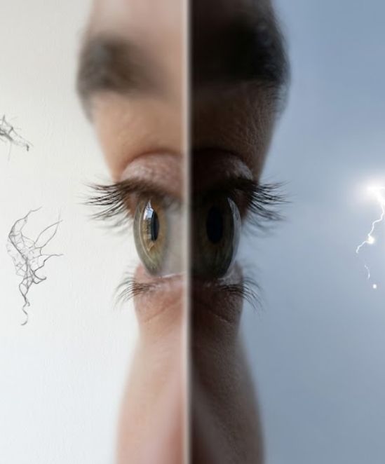
These look like tiny specks, dots, or “cobwebs” drifting in your field of vision. They are actually small clumps of gel casting shadows on your retina. They are most noticeable when looking at a plain background like a white wall or blue sky.

Flashes of Light

Flashes look like lightning bolts or flickering lights in your vision. This happens when the vitreous gel pulls on the retina. If the gel pulls hard enough, it can cause a tear, which is a serious medical issue.

Know Your Risk

You are at a higher risk for serious floaters and flashes if you are very nearsighted, have had cataract surgery, or have experienced a past eye injury. Frequent monitoring helps catch issues before they turn into permanent vision loss.

The Four Stages of Vitreous Change

01

Vitreous Syneresis

The gel inside the eye starts to thin out and clump together. You may notice one or two small floaters that come and go. This is a normal part of aging for most adults.

02

Vitreous Pulling

As the gel shrinks, it pulls away from the back of the eye. This often creates flashes of light. Our specialists monitor this closely to make sure the retina stays safely in place.

03

Retinal Tear

If the shrinking gel pulls a hole in the retina, fluid can leak behind it. This is a critical stage. Treatment at this point can prevent the retina from fully detaching and save your vision.

04

Retinal Detachment

This is a major emergency where the retina lifts away from the back of the eye completely. This stage requires urgent surgery to prevent total blindness in the affected eye.

Symptoms of a Retinal Emergency

Most floaters are normal, but certain signs mean you need to see a specialist immediately. Contact our office right away if you notice:

Notice box: If you see a sudden increase in floaters or flashes, do not wait. This is a medical emergency. A dilated exam is the only way for a specialist to check for a tear and protect your vision.

Advanced Diagnostics & Imaging

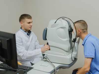
At every visit, our specialists perform a thorough dilated eye exam. We use the most advanced imaging available to check the connection between your vitreous gel and the retina.

Optical Coherence Tomography

This non-invasive test uses light waves to take a cross-section picture of your eye. It allows our doctors to see if the vitreous gel is pulling on the retina or if a small tear is starting to form.

B-Scan Ultrasound
B-Scan Ultrasound

If there is blood in the eye that makes it hard to see the back, we use ultrasound. This uses sound waves to “see” through the eye and check if the retina is still attached and healthy.

Scleral Depression
Scleral Depression

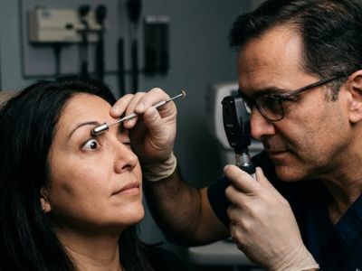
During your exam, the doctor may gently press on the outside of the eye. This allows them to see the very edge of the retina more clearly to ensure no small holes or weak spots are present.

Wide-Field Fundus Photography

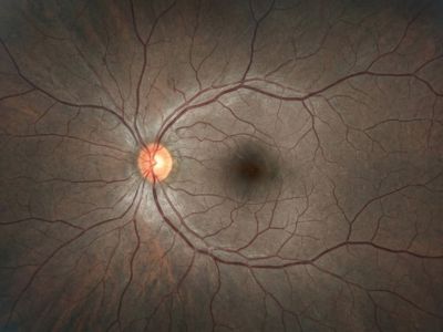
We use specialized cameras to take high-resolution photos of the edges of your retina. This helps us find tiny tears in the far corners of the eye that might be missed during a standard exam.

Dilated Fundus Exam
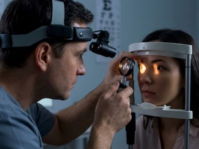
A comprehensive eye examination where pupils are dilated, allowing doctors to carefully inspect retina, vitreous, and optic nerve for abnormalities.

Treatments

The right treatment depends on whether your symptoms are harmless or caused by a tear. Our specialists will review your exam results and explain the best plan for your eye health.

Observation
Observation

If your floaters are caused by normal aging and your retina is healthy, we may simply monitor you. Most people find that floaters become less bothersome over time as the brain learns to ignore them.

Laser therapy
Laser Retinopexy

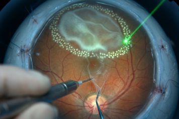
If a retinal tear is found, we use a specialized laser to “weld” the retina back down. This creates a seal around the tear. This prevents fluid from getting under the retina and causing a detachment.

Vitreoretinal Surgery

For advanced cases like a retinal detachment or a large amount of blood in the eye, surgery is needed. Our surgeons perform vitrectomy to repair the retina and clear your vision. RVSU performs over 100 retina surgeries per month.

Our Specialists

All four of our physicians are board-certified ophthalmologists with fellowship training in vitreoretinal surgery. Managing complex retinal tears is a clinical focus for each of them.

Your Eyes Need a Specialist

Sudden flashes and floaters are the primary warning signs of a retinal tear. We are accepting new patients at four locations across Utah. Referrals from primary care physicians and optometrists are always welcome.

Find an RVSU Location Near You

See all of our convenient locations across Utah.