Lattice Degeneration Treatment in Utah

Lattice degeneration is a condition where the edges of your retina become thin and weak. While it often has no symptoms, these thin patches can lead to a retinal tear or detachment if they are not monitored. Our board-certified retina specialists at RVSU use advanced imaging to identify these weak spots and protect your vision. All four of our physicians trained at the number one eye hospital in the country.

Because lattice degeneration happens in the far edges of the eye, you cannot see it without a dilated exam. Our doctors check these areas carefully to decide if you need protective treatment. By catching thinning early, we can often prevent a major eye emergency before it ever starts. Professional care is the best way to manage your retinal detachment risk.

1 in 10 People

in the general population has some degree of lattice degeneration in one or both eyes.

30% of Cases

of retinal detachment are caused by untreated tears in areas of lattice degeneration.

50% More Likely

for patients who are very nearsighted to develop these thin, weak patches in the retina.

What is Lattice Degeneration

What is Lattice Degeneration?

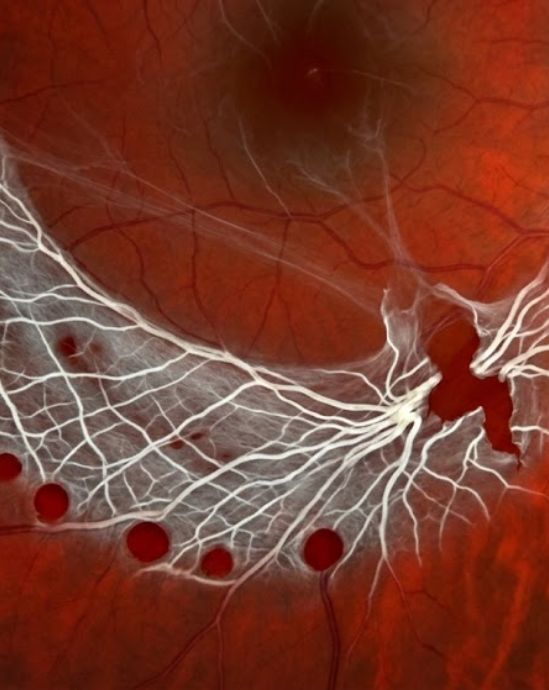
Lattice degeneration gets its name from the “criss-cross” pattern of white lines that appear in the thinned areas of the retina. Over time, the retina loses its thickness and becomes fragile, making it easier for holes or tears to form.

Retinal Thinning

In these specific spots, the retina is much thinner than normal. This thinning usually happens in the peripheral (side) vision. Most people are born with this tendency, and it is often found during a routine eye exam in young adulthood.

Vitreous Attachment

The gel inside your eye, called the vitreous, often sticks more tightly to the edges of these thin patches. When the gel moves or shrinks as you age, it can pull on the thin retina and create a tear.

Know Your Risk

You are at a higher risk for complications if you have a family history of retinal detachment or if you are highly nearsighted. Regular dilated exams are the best way to ensure these weak spots stay stable and do not pull open.

The Four Areas of Impact

01

Stable Thinning

The retina shows small patches of thinning but no holes are present. There is no immediate risk to your vision. Annual monitoring is the standard of care for this stage.

02

Atrophic Holes

Small, round holes form within the thin lattice patches. While these are often stable, they can sometimes allow fluid to leak. More frequent follow-up exams may be needed.

03

Linear Tears

The vitreous gel pulls on the edge of the lattice, creating a sharp tear. This is a higher risk stage that often requires protective laser treatment to prevent a detachment.

04

Retinal Detachment

Fluid gets underneath the retina through a hole or tear, lifting it away from the back of the eye. This is an emergency that requires urgent surgery to save your sight.

Symptoms of a Retinal Tear

Lattice degeneration itself has no symptoms. However, if a thin spot turns into a tear or detachment, you may notice:

Notice box: Lattice degeneration is a “silent” condition. If you are nearsighted or have a family history of eye issues, a dilated exam is the only way to find these thin spots before they cause a tear.

Advanced Diagnostics & Imaging

At every visit, our specialists perform a thorough dilated eye exam. We use the most advanced imaging available to map the thinned areas of your retina.

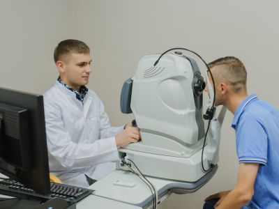
Optical Coherence Tomography
OCT (Optical Coherence Tomography)

This non-invasive test uses light waves to take cross-section pictures of the retina. It helps our doctors measure exactly how thin the lattice patches are and if any fluid is starting to collect.

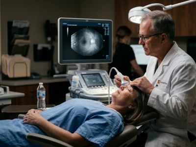
B-Scan Ultrasound
B-Scan Ultrasound

If a cataract or blood blocks the view of the retina, we use sound waves to check the eye. This allows us to see if the retina is still attached to the back of the eye.

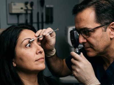
Scleral Depression
Scleral Depression

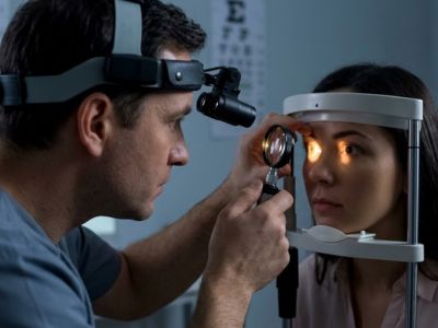
This is the most important part of the exam for lattice. The doctor uses a small tool to gently move the eye so they can see the far edges clearly. This ensures no tiny holes are hidden in the folds of the retina.

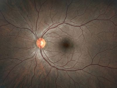
Wide-Field Fundus Photography

Since lattice degeneration happens at the very edge of the eye, we use specialized wide-angle cameras. These photos allow us to track the size and shape of the thinned areas over many years.

Dilated Fundus Exam

Every appointment includes a hands-on clinical exam. Our fellowship-trained physicians personally examine the peripheral retina to ensure your eyes are safe from future tears.

Treatments

Most people with lattice degeneration do not need surgery. Our specialists will review your exam and decide on a plan based on your risk level.

Observation
Observation

If the thin patches are stable and have no holes, we usually monitor them once a year. We will teach you which symptoms to watch for so you know when to call for an urgent checkup.

Laser therapy
Laser Photocoagulation

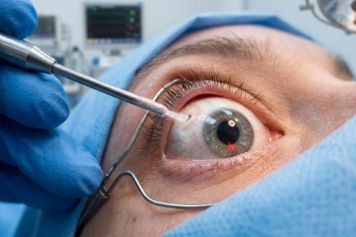
If a hole or tear is found in the lattice, we use a specialized laser to “barrier” the area. This creates a small scar that acts like a weld, keeping the retina firmly attached to the back of the eye.

Cryotherapy

In some cases, we use a freezing probe on the outside of the eye to seal the thin area. This is another way to create a protective seal around a tear to prevent fluid from leaking underneath.

Our Specialists

All four of our physicians are board-certified ophthalmologists with fellowship training in vitreoretinal surgery. Managing peripheral retinal thinning is a clinical focus for each of them.

Your Eyes Need a Specialist

Finding lattice degeneration early is the key to preventing a retinal detachment. We are accepting new patients at four locations across Utah. Referrals from primary care physicians and optometrists are welcome.

Find an RVSU Location Near You

See all of our convenient locations across Utah.