Macular Holes & Macular Puckers Treatment in Utah

The macula is a small spot in the center of your retina. It gives you the sharp vision you need to read, drive, and see faces. Both macular holes and macular puckers affect this area, causing your central vision to become blurry or wavy. Our board-certified retina specialists at RVSU use advanced microsurgery to repair these issues and help you see better.

These conditions often happen because of natural changes in the eye as we get older. Because they only affect your central vision, they do not lead to total blindness, but they can make daily tasks very hard. We provide a clear plan for your macular surgery using the most successful methods available today.

90% Success Rate

for closing macular holes when using modern surgery and following recovery steps.

1 in 10 People

over the age of 60 may develop a macular pucker, though not all will need surgery.

2 to 4 Weeks

is the usual time it takes for vision to begin improving after a successful repair.

What are Macular Holes and Puckers?

While they both affect the same part of the eye, macular holes and puckers are different problems. Both are usually caused by the gel inside your eye pulling on the retina.

Macular Holes

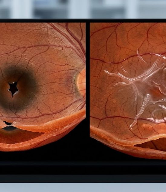
A macular hole is a tiny break in the center of the retina. As the gel inside your eye shrinks, it can pull on the macula until a small hole forms. This causes a dark “blind spot” in the very center of your vision.

Macular Puckers (Epiretinal Membranes)

A macular pucker happens when a thin layer of scar tissue grows over the macula. This tissue can wrinkle or “pucker,” which pulls the retina out of place. This makes straight lines look wavy or bent.

Know Your Risk

Most puckers and holes are caused by aging. However, you may be at a higher risk if you have had a past eye injury, a retinal tear, or if you are very nearsighted. Regular eye exams are the only way to catch these changes early and start eye treatment.

The Four Areas of Impact

01

Vitreous Pulling

The gel inside the eye starts to pull on the center of the retina. You may notice mild blurring in your vision. At this stage, our specialists monitor you closely to see if it gets better on its own.

02

Small Hole Formation

A tiny hole opens in the center of the macula. Your vision may start to look distorted, making it hard to see fine details. Most doctors suggest surgery at this point to stop the hole from getting bigger.

03

Full-Thickness Hole

The hole has opened completely through the layers of the retina. Central vision is mostly blocked by a dark spot. This stage requires surgery to close the hole and protect your sight.

04

Severe Distortion

For puckers, the scar tissue becomes thick and tight. This can cause permanent swelling in the retina. Surgery is needed to “peel” the tissue away and allow the retina to flatten back out.

Symptoms of Macular Issues

Symptoms usually start slowly and only affect one eye at a time. Contact our office right away if you notice any of the following:

Notice box: If you notice that straight lines are looking wavy, you should schedule a dilated exam right away. This is a main sign of a macular issue that needs an eye specialist.

Advanced Diagnostics & Imaging

At every visit, our specialists perform a thorough dilated eye exam. We use the most advanced tools to look at the tiny layers of your macula.

Optical Coherence Tomography
OCT (Optical Coherence Tomography)
This test uses light waves to capture retina images, helping doctors measure macular hole depth and pucker thickness accurately.
Fluorescein Angiography

We may use a special dye to check for leaking fluid. This helps us see if the macular pucker is causing extra swelling that needs to be treated with medicine.

OCTA (OCT Angiography)
A modern, non-invasive imaging technique that maps retinal blood flow without dye, quickly detecting early vascular changes in a comfortable way.
Wide-Field Fundus Photography

We use specialized cameras to take photos of the macula. These photos allow us to track changes over time and plan your surgery.

Dilated Fundus Exam

Every appointment includes a hands-on exam. Our fellowship-trained physicians personally examine your retina to ensure the rest of your eye is healthy.

Treatments

The goal of treatment is to flatten the retina or close the hole to improve your vision. Our specialists will create a plan based on your condition.

Observation
Observation

If a macular pucker is very mild and not bothering your daily life, we may just check it every few months. Some puckers stay the same for years and never need surgery.

Pars Plana Vitrectomy

This is the standard surgery for both holes and puckers. The surgeon removes the vitreous gel to stop it from pulling on the retina. This allows the macular hole to close or the pucker to be fixed.

Membrane Peeling

During surgery for a pucker, the doctor uses tiny tools to gently peel away the thin layer of scar tissue. This allows the retina to relax and return to its normal shape.

Gas Bubble Placement

For macular holes, a temporary gas bubble is placed in the eye to act like a “bandage.” You may need to hold your head in a certain position for a few days to help the hole heal.

Our Specialists

All four of our physicians are board-certified eye doctors with special training in retina surgery. Managing macular hole repair is a main focus for each of them.

Your Eyes Need a Specialist

Macular conditions require very careful surgery to get the best results. We are accepting new patients at four locations across Utah. Referrals from your local eye doctor are welcome.

Find an RVSU Location Near You

See all of our convenient locations across Utah.