Pneumatic Retinopexy

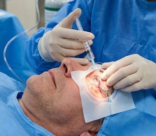
Pneumatic retinopexy is a medical method used to treat certain types of retinal detachment. It is a less invasive option than traditional surgery and is often performed right in our office. During this procedure, a specialist at RVSU injects a small gas bubble into the center of the eye. This bubble floats against the detached area and pushes the retina back against the eye wall. Because it does not require a hospital visit, many patients find this to be a convenient and effective way to restore their vision.

For this treatment to work, the detachment must be located in the upper part of the eye. This allows the gas bubble to float upward and press the retina into place. Our physicians are fellowship-trained experts who carefully evaluate each case to see if this method is the right choice for your eye health.

High Success Rate

When used for the right types of detachments, this method is highly successful at reattaching the retina without major surgery.

Minimal Downtime

Because it is an in-office procedure, patients do not need general anesthesia and can return home immediately afterward.

Localized Treatment

This method allows our doctors to target specific retinal tears with great precision using only a small gas bubble.

Pneumatic Retinopexy

When is Pneumatic Retinopexy Recommended?

A retina specialist may choose this approach if the detachment meets specific medical criteria:

  • Superior Detachments: When the retinal tear or detachment is located in the top half of the eye.
  • Single Tears: This method works best when there is only one tear or a small group of tears close together.
  • Clear Vitreous: The gel inside the eye must be clear enough for the gas bubble to move freely and for the doctor to see the retina.
  • Early Detection: This treatment is most effective when the detachment is caught before it becomes too large or complex.
  • Patient Mobility: The patient must be able to keep their head in a specific position for several days to keep the bubble in place.

How the Gas Bubble Works

The gas bubble stays in the eye for several days or weeks. During this time, it acts as a temporary bandage that holds the retina flat. As the retina stays in contact with the eye wall, the body naturally pumps out the fluid that was trapped behind it. Eventually, the eye replaces the gas bubble with its own natural fluid. Because the gas bubble expands slightly after it is injected, it provides constant, gentle pressure to make sure the retina stays secure while it heals. This process is often combined with laser or freezing treatment to create a permanent seal around the retinal tear.

The Treatment Process

Treating a detached retina is a time-sensitive matter. If you see flashes of light or a shadow in your vision, you should seek a professional exam immediately.

01

Diagnostic Imaging

Our specialists use high-resolution scans to check the size and location of your detachment to ensure it can be fixed with a bubble.

02

Numbing the Eye

We use numbing drops and a small injection to make sure you feel no pain. Most patients say the procedure is very comfortable.

03

Bubble Injection

The doctor uses a very fine needle to place the gas bubble into the eye. You may see a large “spot” in your vision afterward, which is just the bubble floating.

04

Laser or Freezing

Once the retina is pushed back into place, we use a laser or a freezing probe to seal the tear. This ensures the retina stays attached after the bubble is gone.

Post-Procedure Head Positioning

The most important part of this treatment happens at home. To keep the bubble in the right spot, you will need to hold your head in a specific position for most of the day. Your doctor will show you exactly how to tilt your head so the bubble stays pressed against the tear. This is usually required for about one week. You must also avoid lying on your back during this time. Additionally, you cannot fly in an airplane or go to high altitudes until the bubble is fully gone, as the change in pressure can cause the bubble to expand and damage the eye.

Post-Procedure Head Positioning

Our Specialists

All four of our physicians are board-certified ophthalmologists with fellowship training in vitreoretinal surgery.

Retinal Specialists in Utah

The physicians at Retina and Vitreous Surgeons of Utah have extensive experience in performing pneumatic retinopexy. We focus on providing the most effective treatment with the least amount of discomfort for our patients. If you are experiencing symptoms of a retinal detachment, please contact one of our four offices in Murray, Draper, Provo, or Payson for a specialized evaluation.

Find an RVSU Location Near You

See all of our convenient locations across Utah.