
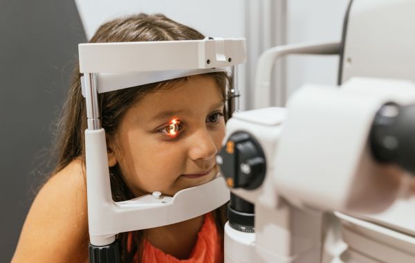
Good vision is an important part of a child’s growth, learning, and everyday life. Children use their vision every day for school, play, and everyday activities. Most common eye issues in children like nearsightedness or a lazy eye are easy to spot and treat. However, some eye conditions happen deep inside the back of the eye, where parents cannot see them. These are called pediatric retinal diseases.
Many children do not realize something is wrong with their vision, so retinal problems may go unnoticed without an eye examination. Understanding how the retina works and recognizing early warning signs can help parents seek treatment before vision problems become more serious.
What Is the Retina and How Does It Work?
To understand retinal disease, it helps to know how the eye processes sight. Think of the eye like a digital camera:
- The Lens: Sits near the front of the eye and focuses light entering through the pupil.
- The Retina: Lines the back wall of the eye like a film or light-sensing screen. It contains millions of tiny cells called photoreceptors.
- The Optic Nerve: Acts like a cable, sending electrical visual signals from the retina straight to the brain.
When light enters your child’s eye, the retina captures that picture and sends it to the brain. If the retina is damaged, swollen, or torn, the image sent to the brain becomes blurry, distorted, or completely blocked.
Because a child’s brain is still developing its visual connections, untreated retinal problems during early years can cause permanent vision loss.
Common Retinal Diseases in Children
Retinal conditions in children are less common than in older adults, but they require specialized medical care when they occur. Here are the main types of pediatric retinal conditions doctors treat:
1. Retinopathy of Prematurity (ROP)
Retinopathy of prematurity affects babies born early usually before 31 weeks of pregnancy or babies who weigh less than 3 pounds at birth.
In premature infants, the blood vessels supplying the retina have not finished growing. After birth, these blood vessels can grow abnormally. Abnormal vessels are fragile; they leak fluid or bleed, creating scar tissue that pulls the retina away from the back of the eye. Early screening in the neonatal intensive care unit (NICU) allows doctors to detect and treat ROP before it harms sight.
2. Pediatric Retinal Detachment
A retinal detachment happens when the retina pulls away from the normal tissue at the back of the eye. In adults, this often happens from aging. In children, retinal detachment is usually caused by:
- Direct eye trauma from sports or accidents
- High nearsightedness (extreme myopia)
- Previous eye surgeries
- Genetic conditions like Marfan syndrome or Stickler syndrome
If the retina separates, its blood supply drops. Without immediate medical care, this can cause quick and permanent vision loss.
3. Retinoblastoma
Retinoblastoma is a rare form of eye cancer that starts in the retina. It mostly affects children under the age of five.
The most noticeable sign is a white glow or reflection in the center of the child’s pupil, often seen in flash photographs. Instead of the typical red-eye look in pictures, the pupil looks bright white, similar to a cat’s eye reflecting light in the dark.
4. Genetic and Inherited Retinal Diseases
Some retinal conditions pass down through family genes. These conditions cause gradual breakdown of the light-sensing cells in the retina:
- Stargardt Disease: A genetic form of macular degeneration that affects central vision, making reading and recognizing faces hard for young kids.
- Retinitis Pigmentosa (RP): A condition that starts by damaging night vision and side vision (peripheral vision), making it hard to see in dim light.
- Coat’s Disease: A rare condition usually affecting one eye in young boys where abnormal blood vessels leak fluid behind the retina, leading to swelling or fluid buildup.
Warning Signs to Watch For
Children may not recognize that their vision has changed, so they often do not mention any problems. Watching for changes in your child’s eyes or daily habits can help identify vision problems sooner.
| Category | What You Might See | Potential Meaning |
|---|---|---|
| Visual Appearance | White reflection in pupil (leukocoria), eyes drifting outward or inward (strabismus), unusual eye shaking (nystagmus) | Possible Retinoblastoma, advanced ROP, or cataracts |
| Physical Habits | Squinting, tilting head constantly, sitting very close to TV or holding books right up to face | Reduced central vision or severe refractive errors |
| Daily Behavior | Frequent tripping, clumsiness in low light, fear of entering dark rooms | Impaired peripheral vision or night blindness (RP) |
| Physical Discomfort | Eye rubbing, sensitivity to normal light (photophobia), frequent headaches | Eye pressure changes or internal eye inflammation |
If your child shows any of these signs, arrange an eye examination with an eye specialist as soon as possible.
Diagnosing Retinal Conditions in Children
Examining a child’s eyes requires specialized equipment and gentle techniques. Pediatric retina specialists use child-friendly tests to get a clear view of the retina:
- Dilated Eye Examination: Doctors use special eye drops to enlarge (dilate) your child’s pupils. This gives the doctor a wide view into the back of the eye using a bright light and a head-mounted magnifying glass called an indirect ophthalmoscope.
- Optical Coherence Tomography (OCT): An OCT scan acts like an ultrasound, but uses safe light waves to capture detailed, cross-sectional pictures of the retina. It shows every layer of the retina and detects tiny amounts of fluid, swelling, or structural damage without touching the eye.
- Wide-Field Retinal Imaging: Special digital cameras take high-resolution, wide-angle photos of the retina in seconds. These pictures help doctors track tiny changes in blood vessels over time.
- Eye Ultrasound (B-Scan): If fluid, cataracts, or blood block the view of the back of the eye, an eye ultrasound uses sound waves to map the retina safely and comfortably.
Retinal Disease Treatments
Treatment depends on the type of retinal disease, its severity, and your child’s overall eye health. Advanced eye care offers several safe treatments:
- Laser and Cryotherapy: Laser energy or cold probes (cryotherapy) treat conditions like Retinopathy of Prematurity and retinal tears. The laser seals leaking blood vessels or glues the edges of a torn retina back into place, preventing full detachment.
- Anti-VEGF Injections: Certain medications stop the body from making a protein called VEGF, which causes abnormal blood vessels to grow and leak. Doctors inject tiny amounts of anti-VEGF medicine directly into the eye under comfortable anesthesia to slow down disease progression.
- Vitrectomy Surgery: During a vitrectomy, a retina surgeon carefully removes the clear gel (vitreous) filling the inside of the eye. This lets the doctor clear out scar tissue, drain accumulated fluid, and repair a detached retina.
- Scleral Buckle: To fix a detached retina, surgeons sometimes place a tiny silicone band (scleral buckle) around the outside of the eye. This band gently pushes the outer wall of the eye against the detached retina until it reattaches and heals.
Protecting Your Child's Eyesight
While you cannot prevent inherited genetic conditions, you can protect your child’s eyes from injury and catch potential diseases early.
1. Schedule Regular Eye Exams
Infants should have their first eye screening around 6 months of age, followed by another exam at age 3 and before starting kindergarten. Children with risk factors such as premature birth or a family history of eye disease need regular visits with a pediatric retina specialist.
2. Protect Eyes During Sports
Eye injuries cause many cases of retinal detachment in school-aged children. Make sure your child wears shatterproof polycarbonate safety goggles when playing sports like baseball, basketball, racquetball, or hockey.
3. Check Flash Photos
When taking flash photos, look at your child’s eyes. If one pupil reflects bright white, yellow, or clear while the other is red, book an eye exam right away.
4. Support Healthy Habits
A balanced diet rich in leafy green vegetables, eggs, and colorful fruits supplies essential nutrients like lutein, zeaxanthin, and omega-3 fatty acids that strengthen retina cells.
Conclusion
Many retinal diseases in children can be managed more successfully when they are diagnosed early. Regular eye examinations, awareness of warning signs, and prompt treatment can help protect your child’s vision and reduce the risk of permanent damage. If you have concerns about your child’s eyesight, an evaluation by a retina specialist can provide answers and the right treatment plan.
At Utah Retina, our board-certified retina specialists are dedicated to providing expert care for children with retinal conditions in a compassionate and supportive environment. Whether your child needs a retinal evaluation, ongoing monitoring, or specialized treatment, our team is here to help. Contact Us today to schedule an appointment and learn how we can help protect your child’s vision.