
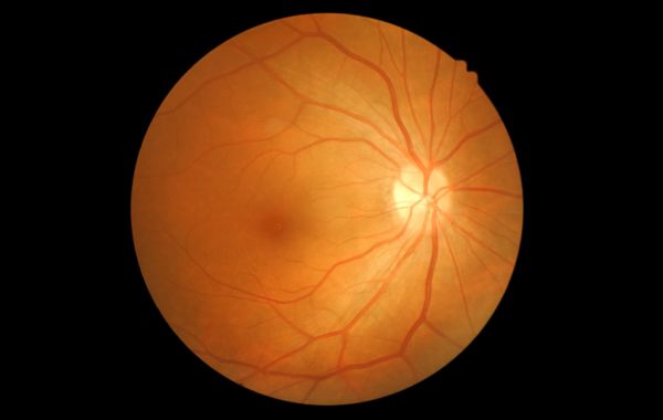
Most people have never heard of retinal vein occlusion until it happens to them or someone in their family. Vision can seem fine one day and blurry the next, with no warning and usually no pain.
Retinal vein occlusion, or RVO, is one of the more common causes of sudden vision loss in adults over 50. It doesn’t get talked about as much as cataracts or glaucoma, but it affects millions of people worldwide. If you or someone in your family has just been told they have this condition, it helps to know what’s going on inside the eye, why it happens, and what your options are.
What Is Retinal Vein Occlusion?
Picture the retina like a camera film at the back of your eye. It needs a steady supply of blood, flowing in through small arteries and out through veins. When one of those veins gets blocked, blood can’t drain properly, and pressure builds up inside it.
Blood and fluid start to leak into the retinal tissue around the blockage, leading to swelling, bleeding, and sometimes areas of the retina that don’t get enough oxygen. The result is blurry vision, dark spots, or in more serious cases, a big chunk of vision disappearing from one eye almost overnight.
There are two main types of RVO, depending on where the blockage happens.
- Central retinal vein occlusion (CRVO) happens when the main vein that drains the whole retina gets blocked, usually causing more widespread vision problems.
- Branch retinal vein occlusion (BRVO) happens when one of the smaller branch veins gets blocked, so the vision loss is usually limited to one area.
BRVO is more common than CRVO and tends to have a somewhat better outlook for vision recovery, though this varies from person to person.
What Causes Retinal Vein Occlusion?
RVO doesn’t happen randomly. There’s almost always something going on in the body that made the blood vessel more likely to clog, and a lot of the same things that raise your risk for heart disease raise your risk for RVO.
Main risk factors doctors look for:
- High blood pressure: The single biggest risk factor. Years of high pressure wear down blood vessel walls, including the small ones in your eyes.
- Diabetes: High blood sugar damages blood vessels and makes blood more likely to clot.
- High cholesterol: Fatty deposits narrow the space blood has to move through.
- Glaucoma: Extra pressure inside the eye can squeeze the retinal vein where it crosses an artery, a common spot for blockages.
- Age: RVO shows up far more often after 50.
- Blood clotting disorders: Some people’s blood clots more easily than it should, seen more often in younger patients with RVO.
- Smoking: Damages blood vessels throughout the body.
- Obesity and inactivity: These tend to feed into the other risk factors above.
In a small number of cases, doctors never pin down a clear cause. That happens more often in younger patients who don’t have any of the usual risk factors.
Symptoms of Retinal Vein Occlusion
Symptoms can show up fast or creep in slowly, depending on how much of the retina is affected. Most people describe it as painless, which is part of what makes it easy to brush off at first.
Common symptoms include:
- Sudden or gradual blurring of vision in one eye
- A dark area or shadow in part of your visual field
- Floaters, small specks or squiggly lines drifting across your view
- Distorted vision, where straight lines look wavy
- In severe cases, a sudden, significant loss of vision in one eye
RVO almost always affects just one eye, so a change in one eye with the other feeling fine is itself a clue that points toward RVO. Because there’s no pain, a lot of people put off getting checked, which isn’t a good idea here. Any sudden vision change is worth a same day or next day visit with an eye doctor.
How Is Retinal Vein Occlusion Diagnosed?
Diagnosis usually starts with a dilated eye exam, where drops widen your pupil so the doctor can get a good look at the back of your eye. A retina specialist checks for telltale signs like bleeding, swelling, and dilated, twisted veins. Some of what shows up can look similar to other retinal conditions, including macular degeneration, which is part of why a full exam matters.
Most patients also get a couple of extra tests. One is called OCT (optical coherence tomography), a quick scan that takes a detailed picture of the retina’s layers. It’s especially good at showing swelling in the macula, the part of the retina you use for sharp, central vision, like reading or recognizing faces. The other common test is fluorescein angiography. A dye is injected into a vein in your arm, and as it travels through the blood vessels in your eye, a camera takes pictures that show doctors exactly where the blood flow is stuck.
Treatment Options for Retinal Vein Occlusion
There’s no single treatment that fixes RVO, since the blockage itself usually can’t be reversed. Treatment instead focuses on managing what the blockage causes, mainly swelling in the macula and unhealthy new blood vessels trying to grow. Catching it early gives the best chance at keeping vision stable.
| Treatment | What It Does | Typical Use |
|---|---|---|
| Anti-VEGF injections | Blocks a substance that makes blood vessels leak and swell | First choice for swelling caused by RVO |
| Steroid injections or implants | Calms down inflammation and swelling in the retina | Used when anti-VEGF injections aren't enough or not tolerated |
| Laser treatment | Seals off leaking vessels or shrinks unhealthy new ones | Used for swelling in some cases, and to stop new vessel growth |
| Managing underlying conditions | Controls blood pressure, blood sugar, and cholesterol | Ongoing, alongside eye treatment |
Anti-VEGF injections are the go-to treatment for most patients with RVO related swelling. These are intravitreal injections, which just means the medicine is given as a shot right into the eye. The eye is numbed first, and the shot itself is quick. Most patients need repeated shots over months, sometimes longer, since the effect wears off over time.
Laser treatment comes into play if new, fragile blood vessels start growing where they shouldn’t. This is sometimes called neovascularization, but it just means new vessels are forming in response to the eye not getting enough blood flow. These new vessels are weak and prone to bleeding, so laser treatment shrinks them down before they cause bigger problems, like bleeding inside the eye or a retinal detachment, which is when the retina pulls away from the back of the eye.
Can Vision Recover After RVO?
This is the question almost every patient asks, and honestly, it depends. Some people, especially with a mild branch occlusion, recover a good amount of vision over time. Others, particularly with a central occlusion or a lot of swelling before treatment starts, are left with some permanent vision loss even after treatment.
Starting treatment early gives you the best odds. The longer swelling goes untreated, the more damage it does to the delicate cells in the macula, and some of that damage doesn’t come back. That’s the main reason doctors push for quick evaluation any time someone notices sudden vision changes.
Living With RVO
Getting diagnosed isn’t really the end point. It’s more the start of regular visits with your eye doctor, checking how the retina is healing and whether swelling or new blood vessels are coming back. Most patients need injections on a set schedule, spaced out further once things settle down.
RVO is also a bit of a wake-up call about your overall vascular health, not just something happening in the eye. For a lot of people, this diagnosis is what finally pushes them to get their blood pressure or blood sugar under control, which lowers the odds of going through this again, in the same eye or the other one.
Protecting Your Vision Starts With Catching It Early
Retinal vein occlusion is hard to predict, and it can happen even to people who feel like they’re taking good care of themselves. What you can control is how fast you act once something looks off. Knowing your risk factors and getting to a retina specialist quickly, instead of waiting it out, usually makes the biggest difference in how much vision you keep. If you’ve noticed sudden blurriness, dark spots, or distorted vision in one eye, don’t sit on it. Reach out to Utah Retina and get it looked at so we can figure out what’s going on.