Every day, you use your eyes to read, drive, watch your favourite shows, and see the people you care about. Many people believe that if they can read the smallest letters on an eye chart, their eyes are healthy. While a routine eye exam is important, it only shows part of the picture.
The back of your eye contains a thin layer of tissue called the retina. The retina captures light and sends visual information to your brain, allowing you to see. If the retina becomes damaged, vision loss can be permanent. Many retinal diseases develop slowly and may not cause symptoms in the early stages. By the time you notice blurry vision or dark spots, damage may already have occurred.
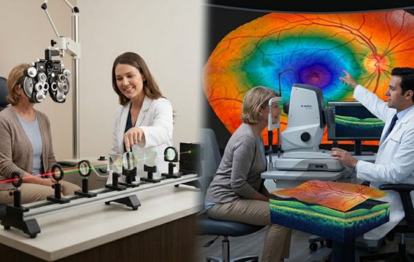
At Utah Retina, advanced imaging technology allows specialists to look deeper into the eye than a routine exam alone. Understanding the difference between a standard eye exam and retinal imaging can help you protect your vision for years to come.
What Happens During a Routine Eye Exam?
A routine eye exam is an important part of maintaining healthy vision. During the visit, your eye care provider checks how well you can see and determines whether you need glasses or contact lenses.
A routine eye exam often includes:
- Visual Acuity Test: You read letters on an eye chart to measure how clearly you can see at different distances.
- Refraction Test: You look through different lenses while the doctor determines which lens provides the clearest vision.
- Eye Muscle and Pressure Tests: Your eye movements are checked, and eye pressure is measured to help screen for conditions such as glaucoma.
- Basic Internal Eye Examination: Using a bright light and special lenses, the doctor examines the front of the eye and takes a quick look at the retina. These tests are excellent for checking vision and overall eye health. However, they have limitations. A routine examination provides only a limited view of the retina, making it more difficult to detect small changes or problems in the outer areas of the eye.
What Is Advanced Retinal Imaging?
Advanced retinal imaging uses specialised technology to create detailed pictures of the inside of your eye. Instead of relying only on a visual examination, doctors can view high-quality digital images of the retina, optic nerve, and blood vessels.
These images help identify changes that may not be visible during a routine exam.
At Utah Retina, specialists commonly use two types of advanced imaging.
1. Ultra-Widefield Digital Photography
Traditional retinal cameras capture only a small portion of the retina at one time. Ultra-widefield imaging provides a much larger view in a single scan.
This technology allows doctors to examine areas near the edges of the retina, where tears, holes, and blood vessel problems can develop. The scan is quick, painless, and does not involve radiation.
2. Optical Coherence Tomography (OCT)
If retinal photography shows the surface of the retina, an OCT scan shows what is happening beneath it.
OCT uses light waves to create highly detailed cross-sectional images of the retina. It allows doctors to examine each retinal layer, measure retinal thickness, and detect fluid or swelling that cannot be seen during a routine examination.
This technology provides valuable information about retinal health and helps identify problems at an early stage.
The Clear Differences at a Glance
| Feature | Routine Eye Exam | Advanced Retinal Imaging (Digital / OCT) |
|---|---|---|
| Primary Goal | Checks vision and determines prescription needs. | Examines retinal structure and tissue health. |
| Field of View | Look at a limited area of the retina. | Captures a wider view of the retina. |
| Layer Analysis | Views the surface of retinal tissue. | Examines individual retinal layers. |
| Permanent Records | Uses written notes in medical records. | Stores detailed digital images for comparison. |
| Early Disease Detection | May detect disease after symptoms appear. | Can identify changes before symptoms develop. |
What Imaging Reveals That Routine Exams May Miss
One of the greatest benefits of retinal imaging is its ability to detect eye diseases before they affect vision. Many serious retinal conditions do not cause pain or noticeable symptoms in their early stages.
Macular Degeneration
Age-related macular degeneration affects the central part of the retina that provides sharp, detailed vision. Before vision becomes blurry, retinal imaging may detect tiny yellow deposits called drusen beneath the retina. Finding these early changes allows doctors to recommend treatments, lifestyle adjustments, or nutritional support that may help slow disease progression.
Diabetic Retinopathy
Diabetes can damage the tiny blood vessels in the retina. Early signs of diabetic retinopathy may be difficult to detect during a routine exam. Advanced imaging can identify small leaks, tiny areas of bleeding, and swelling in the macula before symptoms appear. Early detection helps reduce the risk of serious vision complications.
Glaucoma
Glaucoma is often called the silent thief of sight because it can slowly damage vision without noticeable symptoms. The disease affects the optic nerve, which carries visual information from the eye to the brain. OCT imaging measures the thickness of the nerve fiber layer around the optic nerve. If thinning is detected early, treatment can begin before significant vision loss occurs.
Retinal Tears and Detachments
A retinal tear occurs when the gel inside the eye pulls on the retina and causes it to tear. If fluid passes through the tear, the retina can begin to separate from the back of the eye. This condition, called retinal detachment, is a medical emergency.
Because tears often occur in the outer parts of the retina, they can be difficult to detect during a routine examination. Ultra-widefield imaging helps doctors identify these problems early, allowing treatment before serious vision loss develops.
Why Long-Term Tracking Matters
Retinal images provide more than a snapshot of your eye health today. They also create a record that can be compared over time.
When retinal scans are stored in your medical record, doctors can compare new images with previous ones during future visits.
This side-by-side comparison helps identify even very small changes in retinal tissue, blood vessels, or the optic nerve. Tracking these changes over time allows specialists to detect disease earlier and monitor how well treatments are working.
Because small changes can be difficult to notice during a routine examination alone, long-term imaging plays an important role in protecting vision.
Conclusion
Routine eye exams remain an essential part of eye care. They help evaluate vision, update prescriptions, and identify many common eye conditions. However, advanced retinal imaging provides a much deeper look into the health of your eyes. By capturing detailed images of the retina and its layers, these technologies can detect disease before symptoms appear and help prevent permanent vision loss.
If you have diabetes, a family history of eye disease, or simply want a more complete understanding of your eye health, retinal imaging can provide valuable information and added peace of mind.
Do not wait until vision problems develop. Contact the experienced team at Utah Retina today to schedule a comprehensive retinal evaluation and learn how advanced imaging can help protect your sight.