The human eye allows you to see everything around you, from reading a book to enjoying a beautiful sunset. While many parts of the eye work together to create vision, the retina plays one of the most important roles.
The retina is a thin layer of tissue located at the back of the eye. It captures light, turns it into signals, and sends those signals to the brain. The brain then interprets these signals as the images you see every day.
Understanding retina anatomy can help you learn how vision works and why protecting your retinal health is so important.
What is The Retina?
The retina is a thin, light-sensitive layer of tissue that lines the back of the eye. It works much like the sensor in a camera, capturing light that enters the eye.
Special cells in the retina turn light into signals that travel through the optic nerve to the brain. The brain processes these signals and creates the images you see.
Without a healthy retina, clear vision would not be possible.
Key Functions of the Retina
The retina does much more than simply receive light. It helps process visual information before sending it to the brain.
1. Converting Light into Signals
One of the retina’s main jobs is to turn light into signals that the brain can understand. When light reaches the retina, special cells react to it and create signals that travel to the brain through the optic nerve.
2. Processing Visual Information
The retina begins organising visual information before it reaches the brain. It helps sort details such as brightness, colour, and movement so the brain can process images more efficiently.
3. Improving Contrast and Detail
The retina helps you see differences between light and dark areas. This makes it easier to recognise shapes, read text, and see fine details clearly.
4. Supporting Day and Night Vision
The retina helps you see in different lighting conditions.
- In bright light, it helps you see sharp details and colours.
- In dim light, it helps you see shapes and movement, making it easier to navigate in darker environments.
The Center of Vision: The Macula and Fovea
Two important areas of the retina are responsible for your central vision.
The Macula: The macula is a small area near the centre of the retina. It provides the sharp, detailed vision needed for activities such as reading, driving, and recognising faces.
The Fovea: The fovea is located in the centre of the macula. It contains the highest concentration of cone cells and provides the sharpest vision in the eye.
The rest of the retina supports peripheral vision, which allows you to see objects outside your direct line of sight and helps you move safely through your surroundings.
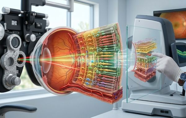
Microscopic Anatomy: The 10 Layers of the Retina
Although the retina is very thin, it is made up of 10 different layers. Each layer has a specific role in helping the retina function properly and support clear vision.
1. Retinal Pigment Epithelium (RPE)
The retinal pigment epithelium, or RPE, is the outermost layer of the retina and sits next to the blood vessel layer beneath it. This layer helps nourish retinal cells, removes waste materials, and absorbs excess light that could interfere with clear vision.
2. Photoreceptor Layer
The photoreceptor layer contains the light-sensitive cells that begin the process of vision. It includes rods, which help with night and peripheral vision, and cones, which allow you to see colours and fine details in bright light.
3. External Limiting Membrane (ELM)
The external limiting membrane helps keep retinal cells properly aligned and supported. Although it is very thin, it plays an important role in maintaining the structure of the retina.
4. Outer Nuclear Layer (ONL)
The outer nuclear layer contains the main cell bodies of the rods and cones. It houses important parts of these light-sensitive cells and helps them remain healthy and function properly.
5. Outer Plexiform Layer (OPL)
The outer plexiform layer is where photoreceptor cells connect with other retinal cells. This connection allows visual signals to move through the retina and supports communication between different cell types.
6. Inner Nuclear Layer (INL)
The inner nuclear layer contains several types of cells that help process visual information. These cells organise signals, support communication throughout the retina, and help maintain a healthy retinal environment.
7. Inner Plexiform Layer (IPL)
The inner plexiform layer serves as another important connection area within the retina. It allows retinal cells to pass signals to ganglion cells, helping visual information continue its journey towards the brain.
8. Ganglion Cell Layer (GCL)
The ganglion cell layer contains ganglion cells, which collect visual information from other retinal cells. These cells help organise processed information and prepare it to be sent to the brain.
9. Nerve Fiber Layer (NFL)
The nerve fibre layer is made up of fibres that extend from ganglion cells. These fibres carry visual signals across the retina and direct them towards the optic nerve, which sends the information to the brain.
10. Internal Limiting Membrane (ILM)
The internal limiting membrane is the innermost layer of the retina. It forms a protective boundary between the retina and the vitreous gel inside the eye, helping maintain the retina’s stability and overall structure.
Conclusion
The retina is a remarkable part of the eye that plays a vital role in vision. Its 10 layers work together to capture light, process visual information, and send signals to the brain. From the retinal pigment epithelium to the nerve fibre layer, each layer helps support healthy eyesight.
Regular eye exams can help detect retinal healthy early and protect your long-term vision. If you notice changes in your central vision or want to learn more about your retinal health, contact Retina and Vitreous Surgeons of Utah today. Contact us to schedule a comprehensive retinal evaluation at one of our convenient locations in Murray, Draper, Provo, or Payson.