
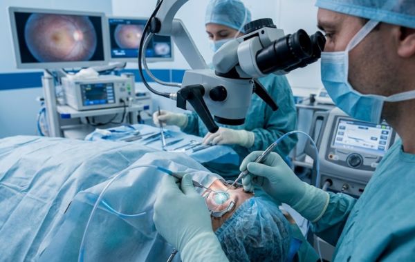
A retinal detachment can be a serious eye problem that needs quick treatment. It happens when the retina, the light-sensitive layer at the back of the eye, pulls away from the tissue that supports it. When this happens, the retina cannot work as it should, which can lead to vision loss if treatment is delayed.
Hearing that you may need retinal detachment surgery can feel stressful. Many patients want to know what happens during the procedure, how the retina is repaired, and what recovery may be like.
The good news is that there are several ways to treat a retinal detachment. The treatment your retina specialist chooses will depend on the type, location, and severity of the detachment.
What Is Retinal Detachment Surgery?
Retinal detachment surgery is used to put the retina back into its normal position and help prevent further vision loss.
A retina specialist first examines the eye and looks at the location and size of the retinal tear or detachment. Based on these findings, the doctor may recommend a procedure such as pneumatic retinopexy, scleral buckle surgery, vitrectomy, or laser treatment.
RVSU offers several treatment options for retinal detachment, including laser surgery or cryopexy, pneumatic retinopexy, scleral buckle, and vitrectomy.
Why Does a Retina Become Detached?
There are several reasons why a retinal detachment may happen.
One common cause is a change in the vitreous, the clear gel that fills the inside of the eye. As we get older, this gel can shrink and pull away from the retina. This is known as a posterior vitreous detachment, or PVD.
Sometimes, this pulling can create a tear in the retina. Fluid can then pass through the tear and collect underneath the retina, causing it to separate from the tissue beneath it.
Other risk factors can include:
- A previous retinal tear
- A strong blow or injury to the eye
- Severe nearsightedness
- Previous eye surgery
- A family history of retinal detachment
- Certain health conditions
What Are the Warning Signs?
Knowing the warning signs of retinal detachment is important because early treatment can help protect vision.
Common symptoms include:
- A sudden increase in floaters
- Flashes of light
- A dark shadow in your vision
- A curtain-like area covering part of your sight
- Sudden blurry or distorted vision
RVSU notes that a sudden increase in floaters, flashes, a shadow or curtain, and sudden changes in vision can be signs of retinal detachment.
If you notice these symptoms, do not wait for them to go away on their own. Contact an eye care professional or seek urgent medical attention.
What Happens Before Retinal Detachment Surgery?
Before treatment, your retina specialist will examine your eye carefully.
The doctor will look for the location of the retinal tear or detachment and check how much of the retina is affected. This helps the doctor decide which treatment is suitable.
Your doctor may also ask about:
- When your symptoms started
- Any recent changes in your vision
- Previous eye surgery
- Any eye injuries
- Your general eye health
- Other health conditions
The goal is to understand the problem clearly before choosing a treatment.
What Happens During Retinal Detachment Treatment?
There is not just one type of retinal detachment surgery. Different procedures are used for different types of retinal problems.
Laser Surgery or Cryopexy
Laser treatment or cryopexy may be used around a retinal tear.
The purpose is to create a small area of scar tissue around the tear. This helps seal the area and can prevent fluid from moving through the tear and causing further retinal detachment.
Laser treatment uses light energy, while cryopexy uses controlled freezing to create the needed scar.
These treatments may sometimes be used with other retinal procedures.
Pneumatic Retinopexy
Pneumatic retinopexy uses a small gas bubble inside the eye.
The doctor places the gas bubble into the eye. The bubble moves against the detached part of the retina and helps push it back into position.
The retinal tear is then treated so that it can seal while the retina heals.
After this procedure, your doctor may give you specific instructions about how to position your head. The position helps keep the gas bubble against the area that needs treatment.
RVSU lists pneumatic retinopexy as one of its treatment options for retinal detachment.
Scleral Buckle Surgery
A scleral buckle is another way to treat a retinal detachment.
During this procedure, a small band is placed around the outside of the eye. The band gently pushes the wall of the eye inward.
This helps reduce the pulling force on the retina and supports the area where the retinal tear occurred.
The buckle usually stays around the eye after surgery.
RVSU describes a scleral buckle as a small band placed around the eye to indent the sclera and reduce traction on the retina.
Vitrectomy
A vitrectomy is another surgical option for retinal detachment.
During this procedure, the surgeon removes some or all of the vitreous gel from inside the eye. Removing the gel can help the surgeon reach the retina and treat the retinal problem.
The surgeon may then use another method, such as a gas bubble, to help hold the retina in place while it heals.
The exact steps can vary depending on the condition of the retina.
Which Retinal Detachment Treatment Will You Need?
There is no single procedure that is right for every patient.
Your retina specialist will consider several factors, including:
| Factor | Why It Matters |
|---|---|
| Location of the tear | Helps the doctor choose the right treatment |
| Size of the detachment | Larger detachments may need surgery |
| Number of retinal tears | Multiple tears can affect the treatment plan |
| Location of the macula | The center of the retina can affect vision |
| Previous eye surgery | May affect which procedure is suitable |
| Overall eye condition | Helps the doctor plan safe treatment |
Your doctor will explain the recommended procedure and why it is being used for your eye.
What Happens After Surgery?
Recovery depends on the type of treatment you receive.
You may have some temporary discomfort, redness, or blurry vision after treatment. Your doctor will provide instructions for caring for your eye during recovery.
If a gas bubble has been placed inside the eye, you may need to keep your head in a specific position for a period of time. Following these instructions is important because the bubble needs to remain in the right position while the retina heals.
You may also need to use prescribed eye drops and attend follow-up appointments.
Your retina specialist will monitor your eye and check how the retina is healing.
Can Vision Return After Retinal Detachment Surgery?
The amount of vision recovery varies from person to person.
Several factors can affect the result, including how quickly the detachment was treated, how much of the retina was affected, and whether the macula was involved.
Retinal detachment is considered a serious eye condition because the retina can lose its ability to function normally when it is separated from its supporting tissue. Prompt diagnosis and treatment are important for protecting vision.
Even after successful surgery, vision may take time to improve. Some patients may continue to have changes in their vision during recovery.
What Should You Avoid After Surgery?
Your retina specialist will give you specific instructions based on your procedure.
Depending on your treatment, you may need to:
- Avoid heavy activity for a period of time
- Use prescribed eye drops
- Avoid rubbing or pressing on the eye
- Attend all follow-up visits
- Follow any head-positioning instructions
- Tell other doctors if you have a gas bubble in your eye
If you have a gas bubble, your doctor may also give you special instructions about air travel and certain types of anesthesia. Always tell another doctor about the gas bubble before having any medical procedure.
When Should You Seek Help After Surgery?
Some discomfort and changes in vision can happen during recovery, but new or worsening symptoms should be reported.
Contact your eye care team promptly if you notice:
- Sudden loss of vision
- A major increase in floaters
- New flashes of light
- Increasing pain
- Severe redness
- A new dark curtain or shadow
- Other sudden changes in your vision
Your doctor can determine whether these symptoms are part of normal recovery or need further treatment.
Final Thoughts
Retinal detachment surgery is designed to repair the retina and help protect your vision. Depending on your condition, treatment may involve laser or cryopexy, pneumatic retinopexy, scleral buckle surgery, or vitrectomy.
The right treatment depends on the location and severity of the retinal detachment and the overall condition of your eye. RVSU provides several retinal detachment treatment options and recommends prompt medical attention when warning signs appear.
If you experience sudden flashes, many new floaters, a dark curtain, or sudden changes in vision, seek immediate eye care. Early treatment can be an important step in protecting your sight.