
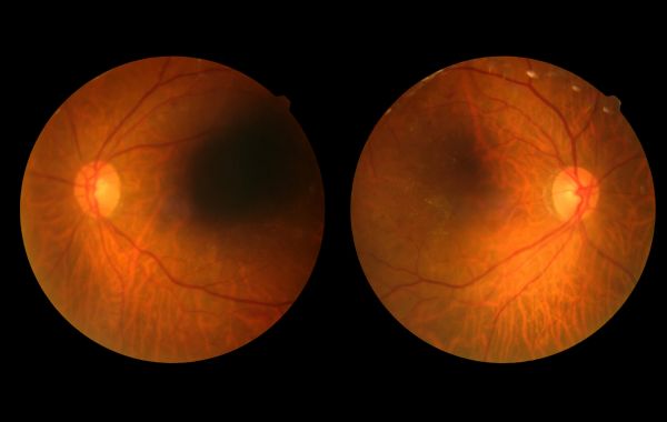
Learning that you have diabetic retinopathy can be scary. You may worry about your eyesight and daily life. Finding it early is the first step to protecting your vision. Your retina specialist will work with you and your doctor to create a treatment plan that helps protect your vision.
With early treatment and good blood sugar control, many people keep their vision. Whether you were recently diagnosed or are seeking specialized care, this guide breaks down what diabetic retinopathy is, how it affects your eyes, available treatments, and five practical steps you can take today to protect your sight.
What Is Diabetic Retinopathy?
Diabetic retinopathy is an eye condition caused by diabetes. It affects the retina, which is the light-sensitive layer of tissue at the back of your eye.The retina works like a camera. It captures light and sends signals to your brain so you can see.
High blood sugar over time damages the tiny blood vessels in the retina. These weak vessels can:
- Leak fluid or blood into the retina.
- Swell and close off, stopping healthy blood flow.
- Cause new, abnormal blood vessels to grow on the surface of the retina.
If left untreated, these changes can cause permanent vision loss or blindness.
Stages of Diabetic Retinopathy
Diabetic retinopathy usually gets worse slowly. Doctors divide it into two stages: non-proliferative (early) and proliferative (advanced).
1. Non-Proliferative Diabetic Retinopathy (NPDR)
This is the early stage of diabetic eye disease. Most people do not notice vision changes during this early stage. Inside the eye, tiny blood vessels swell and form tiny pouches called microaneurysms. These weak spots can leak fluid into the retina.
As NPDR progresses:
- Blood vessels become blocked.
- Parts of the retina stop getting enough oxygen.
- The macula, the part of the retina that gives you clear vision, can swell. This is called diabetic macular edema (DME).
2. Proliferative Diabetic Retinopathy (PDR)
PDR is the more severe stage of the disease. Because many blood vessels in the retina are blocked, the eye tries to grow new blood vessels to replace them. This is called the growth of new blood vessels.
Unfortunately, these new blood vessels are fragile and do not grow properly. They easily leak blood into the center of the eye, causing severe vision blurriness or dark spots. These abnormal vessels can also create scar tissue. As the scar tissue shrinks, it can pull the retina away from the back of the eye, causing a retinal detachment.
Symptoms to Watch For
In the early stages, diabetic retinopathy often has no symptoms at all. You may not notice any symptoms at first. Your vision may seem normal. This is why annual dilated eye exams are essential for anyone living with diabetes.
As the disease progresses, you may notice:
- Blurry or double vision
- Floating spots, dark strings, or cobweb-like shapes (floaters)
- Fluctuating vision (vision that changes from day to day)
- Poor night vision
- Blank or dark areas in your field of vision
- Colors looking faded or washed out
If you experience sudden vision changes or a sudden increase in floaters, contact a retina specialist immediately.
How Doctors Diagnose Diabetic Retinopathy
A regular vision test cannot fully check your retina. A retina specialist uses specialized diagnostic tools to look at the blood vessels in the back of your eye.
Here are the main tests used during an evaluation:
| Diagnostic Test | What It Does | Why It Helps |
|---|---|---|
| Dilated Eye Exam | Special eye drops widen your pupils so the doctor can view the retina clearly. | Allows the specialist to check for leaking blood vessels, swelling, and tissue damage. |
| Optical Coherence Tomography (OCT) | A non-invasive scan that takes detailed cross-section images of your retina. | Measures retina thickness and detects tiny amounts of fluid caused by diabetic macular edema. |
| Fluorescein Angiography | A harmless dye is injected into your arm, and a camera snaps pictures as it flows through eye vessels. | Highlights blocked blood vessels, severe leaks, and new abnormal vessel growth. |
| Fundus Photography | High-resolution photographs capture detailed images of the back of the eye. | Provides a baseline image to track changes in your retina over time. |
Diabetic Retinopathy Treatments
Your treatment depends on how serious the disease is and whether your retina is swollen. Early stages may only require close monitoring, while advanced stages need active medical treatment.
1. Anti-VEGF Injections
Anti-VEGF injections are a common treatment for diabetic macular edema and advanced diabetic retinopathy. VEGF is a protein that causes unhealthy blood vessels to grow and leak.
Anti-VEGF medications block this protein. They help:
- Reduce swelling in the macula.
- Slow down or reverse abnormal blood vessel growth.
- Improve or stabilize vision.
The doctor numbs your eye with drops before giving the injection. Most patients feel minimal discomfort during the procedure.
2. Laser Photocoagulation
Laser treatment uses a small beam of light to treat damaged parts of the retina. There are two main types of laser treatment:
- Focal Laser Treatment: Targets specific leaking blood vessels in small areas near the macula to slow down fluid leaks.
- Panretinal Photocoagulation (PRP): Treats broader areas of the retina away from the center. This causes abnormal blood vessels to shrink and stops new ones from growing.
3. Steroid Injections and Implants
If anti-VEGF injections do not fully clear retinal swelling, doctors may use corticosteroid injections or tiny slow-release implants placed inside the eye. These medications lower inflammation and reduce swelling in the macula.
4. Vitrectomy Surgery
In advanced cases of proliferative diabetic retinopathy, blood may bleed heavily into the clear gel that fills the middle of the eye (the vitreous). Scar tissue may also pull on the retina.
A vitrectomy is surgery to remove blood and scar tissue from inside the eye. This allows light to reach the retina properly again and lowers the risk of retinal detachment.
5 Steps to Protect Your Vision
Getting a diagnosis is a call to action. Here are five practical steps you can take today to protect your eyesight and keep your vision clear.
Step 1: See a Retina Specialist
Diabetic retinopathy requires specialized medical care. A retina specialist focuses exclusively on diseases of the retina and macula. They have special training and tools to find eye problems early.
Set up a regular exam schedule with your eye doctor and attend every appointment, even if your vision feels fine.
Step 2: Control Your Blood Sugar
High blood sugar is the primary cause of blood vessel damage in your eyes. Keeping your blood sugar under control can slow down the disease.
Work with your doctor to keep track of your HbA1c level. An HbA1c test measures your average blood sugar over the last three months. Even a small drop in your HbA1c can help protect your eyes.
Step 3: Control Blood Pressure and Cholesterol
High blood pressure (hypertension) and high cholesterol place extra strain on fragile blood vessels. When high blood pressure combines with high blood sugar, the risk of retinal bleeding and fluid leakage increases.
- Aim to keep blood pressure at or below your doctor’s recommended target.
- Eat a heart-healthy diet low in saturated fats and sodium.
- Take prescribed medications regularly to control blood pressure and lipid levels.
Step 4: Build Healthy Habits
Small daily decisions add up to big protections for your eyes:
- Quit Smoking: Tobacco use narrows blood vessels and reduces oxygen delivery to eye tissues, accelerating diabetic damage.
- Stay Physically Active: Regular, moderate exercise helps lower blood sugar and improves blood circulation throughout the body. Talk to your doctor before starting heavy weightlifting if you have advanced retinopathy, as straining can raise eye pressure.
- Eat Nutritious Foods: Include leafy green vegetables, citrus fruits, nuts, and fish rich in omega-3 fatty acids in your meals.
Step 5: Check Your Vision at Home
Keep track of changes in your vision between doctor visits.
- Check your vision one eye at a time by covering one eye and looking at a fixed point on a wall or doorway.
- Use an Amsler grid if recommended by your eye doctor. An Amsler grid is a simple pattern of straight lines that helps you notice blurry, wavy, or blank areas in your central vision.
- Seek immediate medical help if you notice sudden dark spots, a dark curtain falling over your field of vision, or sudden blurriness.
Conclusion
A diabetic retinopathy diagnosis can feel scary, but early care can make a big difference. By controlling your blood sugar and seeing your eye doctor regularly, you can help protect your vision.
Your eyesight is priceless, and you do not have to navigate this journey alone. Contact Us today at Utah Retina to schedule a comprehensive retinal examination with our compassionate specialists and build a personalized treatment plan tailored to your needs.